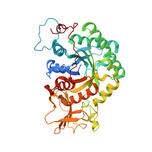
Crystal structure and mutagenesis analysis of chitinase CrChi1 from the nematophagous fungus Clonostachys rosea in complex with the inhibitor caffeine
Yang, J., Gan, Z., Lou, Z., Tao, N., Mi, Q., Liang, L., Sun, Y., Guo, Y., Huang, X., Zou, C., Rao, Z., Meng, Z., Zhang, K.-Q.(2010) Microbiology (N Y) 156: 3566-3574
- PubMed: 20829286
- DOI: https://doi.org/10.1099/mic.0.043653-0
- Primary Citation of Related Structures:
3G6L, 3G6M - PubMed Abstract:
Chitinases are a group of enzymes capable of hydrolysing the β-(1,4)-glycosidic bonds of chitin, an essential component of the fungal cell wall, the shells of nematode eggs, and arthropod exoskeletons. Chitinases from pathogenic fungi have been shown to be putative virulence factors, and can play important roles in infecting hosts. However, very limited information is available on the structure of chitinases from nematophagous fungi. Here, we present the 1.8 Å resolution of the first structure of a Family 18 chitinase from this group of fungi, that of Clonostachys rosea CrChi1, and the 1.6 Å resolution of CrChi1 in complex with a potent inhibitor, caffeine. Like other Family 18 chitinases, CrChi1 has the DXDXE motif at the end of strand β5, with Glu174 as the catalytic residue in the middle of the open end of the (β/α)(8) barrel. Two caffeine molecules were shown to bind to CrChi1 in subsites -1 to +1 in the substrate-binding domain. Moreover, site-directed mutagenesis of the amino acid residues forming hydrogen bonds with caffeine molecules suggests that these residues are important for substrate binding and the hydrolytic process. Our results provide a foundation for elucidating the catalytic mechanism of chitinases from nematophagous fungi and for improving the pathogenicity of nematophagous fungi against agricultural pest hosts.
Organizational Affiliation:
Laboratory for Conservation and Utilization of Bio-Resources, and Key Laboratory for Microbial Resources of the Ministry of Education, Yunnan University, Kunming 650091, PR China.


