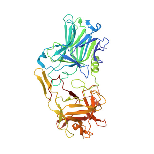
Glycosylated SV2 and gangliosides as dual receptors for botulinum neurotoxin serotype F
Fu, Z., Chen, C., Barbieri, J.T., Kim, J.J., Baldwin, M.R.(2009) Biochemistry 48: 5631-5641
- PubMed: 19476346
- DOI: https://doi.org/10.1021/bi9002138
- Primary Citation of Related Structures:
3FUO, 3FUQ - PubMed Abstract:
Botulinum neurotoxin causes rapid flaccid paralysis through the inhibition of acetylcholine release at the neuromuscular junction. The seven BoNT serotypes (A-G) have been proposed to bind motor neurons via ganglioside-protein dual receptors. To date, the structure-function properties of BoNT/F host receptor interactions have not been resolved. Here, we report the crystal structures of the receptor binding domains (HCR) of BoNT/A and BoNT/F and the characterization of the dual receptors for BoNT/F. The overall polypeptide fold of HCR/A is essentially identical to the receptor binding domain of the BoNT/A holotoxin, and the structure of HCR/F is very similar to that of HCR/A, except for two regions implicated in neuronal binding. Solid phase array analysis identified two HCR/F binding glycans: ganglioside GD1a and oligosaccharides containing an N-acetyllactosamine core. Using affinity chromatography, HCR/F bound native synaptic vesicle glycoproteins as part of a protein complex. Deglycosylation of glycoproteins using alpha(1-3,4)-fucosidase, endo-beta-galactosidase, and PNGase F disrupted the interaction with HCR/F, while the binding of HCR/B to its cognate receptor, synaptotagmin I, was unaffected. These data indicate that the HCR/F binds synaptic vesicle glycoproteins through the keratan sulfate moiety of SV2. The interaction of HCR/F with gangliosides was also investigated. HCR/F bound specifically to gangliosides that contain alpha2,3-linked sialic acid on the terminal galactose of a neutral saccharide core (binding order GT1b = GD1a >> GM3; no binding to GD1b and GM1a). Mutations within the putative ganglioside binding pocket of HCR/F decreased binding to gangliosides, synaptic vesicle protein complexes, and primary rat hippocampal neurons. Thus, BoNT/F neuronal discrimination involves the recognition of ganglioside and protein (glycosylated SV2) carbohydrate moieties, providing a structural basis for the high affinity and specificity of BoNT/F for neurons.
Organizational Affiliation:
Department of Biochemistry, Medical Collegeof Wisconsin, 8701 Watertown Plank Road, Milwaukee, Wisconsin 53226, USA.