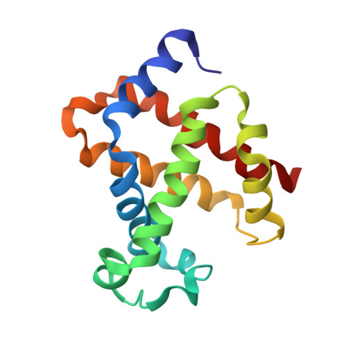
3EU1

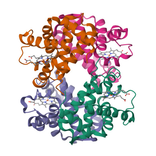
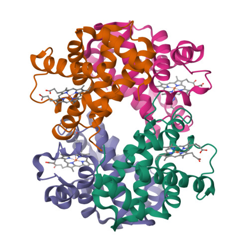
Crystal Structure determination of goat hemoglobin (Capra hircus) at 3 angstrom resolution


Experimental Data Snapshot

  • Method: X-RAY DIFFRACTION
  • Resolution: 3.00 Å
  • R-Value Free: 0.278 
  • R-Value Work: 0.213 
  • R-Value Observed: 0.219 

Starting Model: experimental
View more details

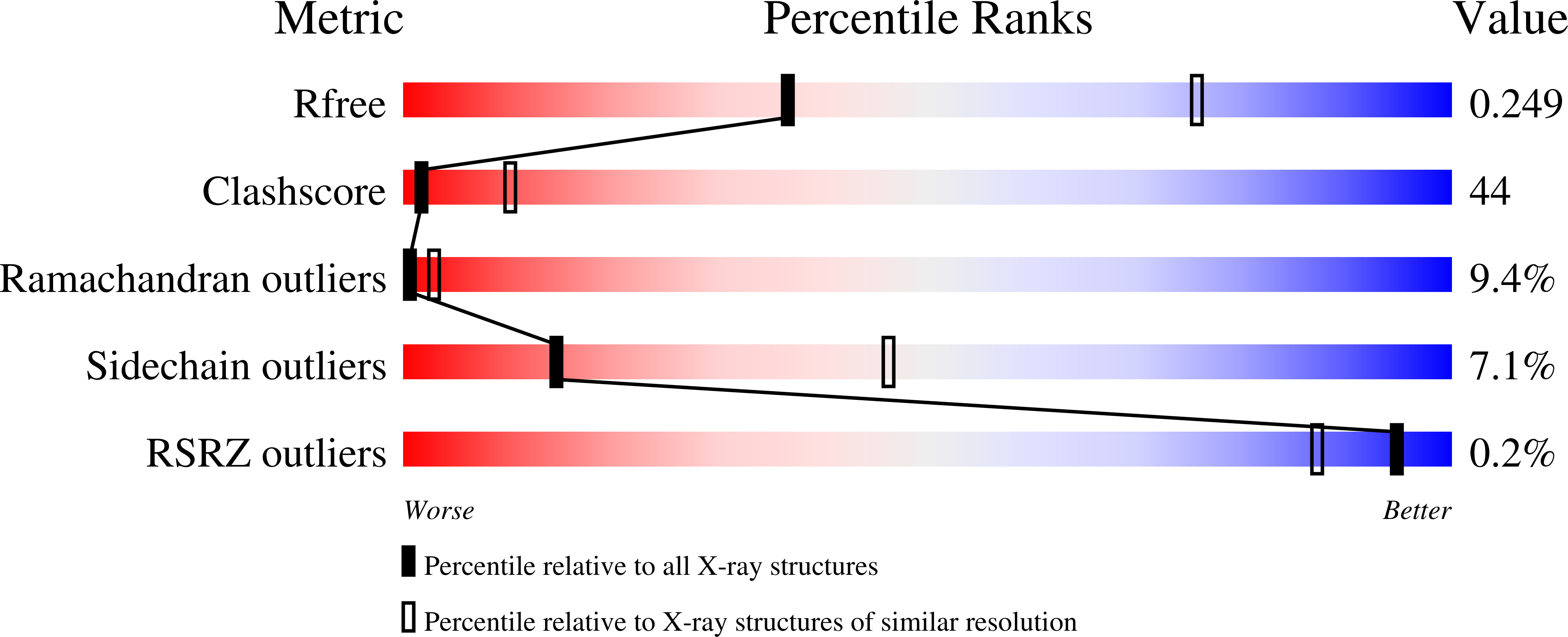
wwPDB Validation   3D Report Full Report


Ligand Structure Quality Assessment 


This is version 1.3 of the entry. See complete history


Literature

Purification, crystallization and preliminary X-ray diffraction studies on goat (Capra hircus) hemoglobin - a low oxygen affinity species

Sathya Moorthy, P.Neelagandan, K.Balasubramanian, M.Ponnuswamy, M.N.

(2009) Protein Pept Lett 16: 454-456

  • DOI: https://doi.org/10.2174/092986609787847992
  • Primary Citation of Related Structures:  
    3EU1

  • PubMed Abstract: 

    Hemoglobin is a vital protein present in almost all higher species. It is a transport protein involved in carrying oxygen from lungs to tissues and carbon dioxide back to lungs by an intrinsically coordinated manner. Even though a good amount of work has been carried out in this direction there exists scarcity of structural insight on low oxygen affinity species. Attempts are being made to unravel the structural insight of this low oxygen affinity species. Goat blood plasma was collected, treated with EDTA to avoid blood clotting and purification was accomplished using DEAE-anion chromatographic column. The goat hemoglobin was crystallized using 50mM of phosphate buffer at pH 6.7 with 1M NaCl and PEG 3350 as precipitant by hanging drop vapor diffusion method. Crystals obtained are screened and suitable crystals are taken for data collection using mar345dtb as image plate detector system. Goat hemoglobin crystal diffracted up to 2.61 A resolution. Goat hemoglobin crystallizes in orthorhombic space group P212(1)2(1) as a whole biological molecule in the asymmetric unit with cell dimensions a=53.568A, b=67.365A, c=154.183A.


  • Organizational Affiliation

    Centre of Advanced Study in Crystallography and Biophysics, University of Madras, Guindy Campus, Chennai - 600 025, India.


Macromolecules
Find similar proteins by:  (by identity cutoff)  |  3D Structure
Entity ID: 1
MoleculeChains Sequence LengthOrganismDetailsImage
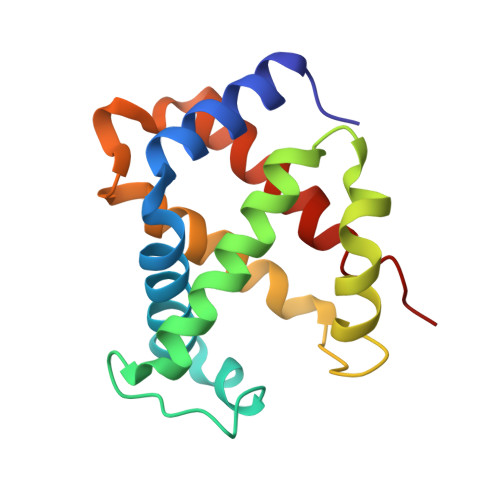
Hemoglobin subunit alpha-1/2
A, C
141Capra hircusMutation(s): 0 
UniProt
Find proteins for P0CH25 (Capra hircus)
Explore P0CH25 
Go to UniProtKB:  P0CH25
Entity Groups  
Sequence Clusters30% Identity50% Identity70% Identity90% Identity95% Identity100% Identity
UniProt GroupP0CH25
Sequence Annotations
Expand
  • Reference Sequence
Find similar proteins by:  (by identity cutoff)  |  3D Structure
Entity ID: 2
MoleculeChains Sequence LengthOrganismDetailsImage
Hemoglobin subunit beta-A
B, D
145Capra hircusMutation(s): 0 
UniProt
Find proteins for P02077 (Capra hircus)
Explore P02077 
Go to UniProtKB:  P02077
Entity Groups  
Sequence Clusters30% Identity50% Identity70% Identity90% Identity95% Identity100% Identity
UniProt GroupP02077
Sequence Annotations
Expand
  • Reference Sequence
Experimental Data & Validation

Experimental Data

  • Method: X-RAY DIFFRACTION
  • Resolution: 3.00 Å
  • R-Value Free: 0.278 
  • R-Value Work: 0.213 
  • R-Value Observed: 0.219 
  • Space Group: P 21 21 21
Unit Cell:
Length ( Å )Angle ( ˚ )
a = 53.678α = 90
b = 66.896β = 90
c = 157.593γ = 90
Software Package:
Software NamePurpose
MAR345data collection
PHASERphasing
REFMACrefinement
AUTOMARdata reduction
SCALEPACKdata scaling

Structure Validation

View Full Validation Report


Ligand Structure Quality Assessment 


Entry History 

Deposition Data

Revision History  (Full details and data files)

  • Version 1.0: 2009-11-10
    Type: Initial release
  • Version 1.1: 2011-07-13
    Changes: Version format compliance
  • Version 1.2: 2018-06-20
    Changes: Data collection, Database references, Structure summary
  • Version 1.3: 2023-11-01
    Changes: Data collection, Database references, Derived calculations, Refinement description