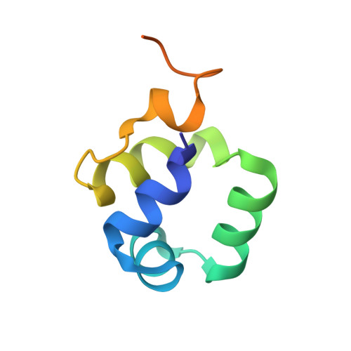
3BD1

Structure of the Cro protein from putative prophage element Xfaso 1 in Xylella fastidiosa strain Ann-1


Experimental Data Snapshot

  • Method: X-RAY DIFFRACTION
  • Resolution: 1.40 Å
  • R-Value Free: 0.182 
  • R-Value Work: 0.147 
  • R-Value Observed: 0.147 

Starting Model: experimental
View more details

wwPDB Validation   3D Report Full Report


This is version 1.2 of the entry. See complete history


Literature

Transitive homology-guided structural studies lead to discovery of Cro proteins with 40% sequence identity but different folds

Roessler, C.G.Hall, B.M.Anderson, W.J.Ingram, W.M.Roberts, S.A.Montfort, W.R.Cordes, M.H.

(2008) Proc Natl Acad Sci U S A 105: 2343-2348

  • DOI: https://doi.org/10.1073/pnas.0711589105
  • Primary Citation of Related Structures:  
    2PIJ, 3BD1

  • PubMed Abstract: 

    Proteins that share common ancestry may differ in structure and function because of divergent evolution of their amino acid sequences. For a typical diverse protein superfamily, the properties of a few scattered members are known from experiment. A satisfying picture of functional and structural evolution in relation to sequence changes, however, may require characterization of a larger, well chosen subset. Here, we employ a "stepping-stone" method, based on transitive homology, to target sequences intermediate between two related proteins with known divergent properties. We apply the approach to the question of how new protein folds can evolve from preexisting folds and, in particular, to an evolutionary change in secondary structure and oligomeric state in the Cro family of bacteriophage transcription factors, initially identified by sequence-structure comparison of distant homologs from phages P22 and lambda. We report crystal structures of two Cro proteins, Xfaso 1 and Pfl 6, with sequences intermediate between those of P22 and lambda. The domains show 40% sequence identity but differ by switching of alpha-helix to beta-sheet in a C-terminal region spanning approximately 25 residues. Sedimentation analysis also suggests a correlation between helix-to-sheet conversion and strengthened dimerization.


  • Organizational Affiliation

    Department of Biochemistry and Molecular Biophysics, University of Arizona, Tucson, AZ 85721, USA.


Macromolecules
Find similar proteins by:  (by identity cutoff)  |  3D Structure
Entity ID: 1
MoleculeChains Sequence LengthOrganismDetailsImage
Cro protein
A, B, C
79Xylella fastidiosa subsp. sandyi Ann-1Mutation(s): 0 
Entity Groups  
Sequence Clusters30% Identity50% Identity70% Identity90% Identity95% Identity100% Identity
Sequence Annotations
Expand
  • Reference Sequence
Small Molecules
Ligands 3 Unique
IDChains Name / Formula / InChI Key2D Diagram3D Interactions
SO4
Query on SO4

Download Ideal Coordinates CCD File 
G [auth C],
I [auth C]
SULFATE ION
O4 S
QAOWNCQODCNURD-UHFFFAOYSA-L
GOL
Query on GOL

Download Ideal Coordinates CCD File 
D [auth A],
E [auth A],
F [auth B],
J [auth C]
GLYCEROL
C3 H8 O3
PEDCQBHIVMGVHV-UHFFFAOYSA-N
CL
Query on CL

Download Ideal Coordinates CCD File 
H [auth C]CHLORIDE ION
Cl
VEXZGXHMUGYJMC-UHFFFAOYSA-M
Experimental Data & Validation

Experimental Data

  • Method: X-RAY DIFFRACTION
  • Resolution: 1.40 Å
  • R-Value Free: 0.182 
  • R-Value Work: 0.147 
  • R-Value Observed: 0.147 
  • Space Group: P 32
Unit Cell:
Length ( Å )Angle ( ˚ )
a = 66.97α = 90
b = 66.97β = 90
c = 54.36γ = 120
Software Package:
Software NamePurpose
d*TREKdata scaling
REFMACrefinement
PDB_EXTRACTdata extraction
XDISPLAYFdata collection
d*TREKdata reduction
ACORNphasing

Structure Validation

View Full Validation Report


Entry History 

Deposition Data

Revision History  (Full details and data files)

  • Version 1.0: 2008-03-25
    Type: Initial release
  • Version 1.1: 2011-07-13
    Changes: Source and taxonomy, Version format compliance
  • Version 1.2: 2023-08-30
    Changes: Data collection, Database references, Derived calculations, Refinement description