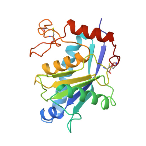
3AIG

ADAMALYSIN II WITH PEPTIDOMIMETIC INHIBITOR POL656


Experimental Data Snapshot

  • Method: X-RAY DIFFRACTION
  • Resolution: 2.80 Å
  • R-Value Work: 0.194 
  • R-Value Observed: 0.194 

Starting Model: experimental
View more details

wwPDB Validation   3D Report Full Report


This is version 2.0 of the entry. See complete history


Literature

Structures of adamalysin II with peptidic inhibitors. Implications for the design of tumor necrosis factor alpha convertase inhibitors.

Gomis-Ruth, F.X.Meyer, E.F.Kress, L.F.Politi, V.

(1998) Protein Sci 7: 283-292

  • DOI: https://doi.org/10.1002/pro.5560070207
  • Primary Citation of Related Structures:  
    2AIG, 3AIG

  • PubMed Abstract: 

    Crotalus adamanteus snake venom adamalysin II is the structural prototype of the adamalysin or ADAM family comprising proteolytic domains of snake venom metalloproteinases, multimodular mammalian reproductive tract proteins, and tumor necrosis factor alpha convertase, TACE, involved in the release of the inflammatory cytokine, TNFalpha. The structure of adamalysin II in noncovalent complex with two small-molecule right-hand side peptidomimetic inhibitors (Pol 647 and Pol 656) has been solved using X-ray diffraction data up to 2.6 and 2.8 A resolution. The inhibitors bind to the S'-side of the proteinase, inserting between two protein segments, establishing a mixed parallel-antiparallel three-stranded beta-sheet and coordinate the central zinc ion in a bidentate manner via their two C-terminal oxygen atoms. The proteinase-inhibitor complexes are described in detail and are compared with other known structures. An adamalysin-based model of the active site of TACE reveals that these small molecules would probably fit into the active site cleft of this latter metalloproteinase, providing a starting model for the rational design of TACE inhibitors.


  • Organizational Affiliation

    Department de Biologia Molecular i Cel.lular, Centre d'Investigació i Desenvolupament C.S.I.C., Barcelona, Spain. xgrcri@cid.csic.es


Macromolecules
Find similar proteins by:  (by identity cutoff)  |  3D Structure
Entity ID: 1
MoleculeChains Sequence LengthOrganismDetailsImage
ADAMALYSIN II202Crotalus adamanteusMutation(s): 0 
EC: 3.4.24.46
UniProt
Find proteins for P34179 (Crotalus adamanteus)
Explore P34179 
Go to UniProtKB:  P34179
Entity Groups  
Sequence Clusters30% Identity50% Identity70% Identity90% Identity95% Identity100% Identity
UniProt GroupP34179
Sequence Annotations
Expand
  • Reference Sequence
Small Molecules
Ligands 4 Unique
IDChains Name / Formula / InChI Key2D Diagram3D Interactions
0ZC
Query on 0ZC

Download Ideal Coordinates CCD File 
E [auth A](3R)-2-[N-(furan-2-ylcarbonyl)-L-leucyl]-2,3,4,9-tetrahydro-1H-beta-carboline-3-carboxylic acid
C23 H25 N3 O5
DNEGAJMITFSILM-HKUYNNGSSA-N
SO4
Query on SO4

Download Ideal Coordinates CCD File 
D [auth A]SULFATE ION
O4 S
QAOWNCQODCNURD-UHFFFAOYSA-L
ZN
Query on ZN

Download Ideal Coordinates CCD File 
B [auth A]ZINC ION
Zn
PTFCDOFLOPIGGS-UHFFFAOYSA-N
CA
Query on CA

Download Ideal Coordinates CCD File 
C [auth A]CALCIUM ION
Ca
BHPQYMZQTOCNFJ-UHFFFAOYSA-N
Modified Residues  1 Unique
IDChains TypeFormula2D DiagramParent
PCA
Query on PCA
A
L-PEPTIDE LINKINGC5 H7 N O3GLN
Biologically Interesting Molecules (External Reference) 1 Unique
Entity ID: 5
IDChains NameType/Class2D Diagram3D Interactions
PRD_000372 (0ZC)
Query on PRD_000372
E [auth A]Furoyl-LeusycloTrp; Po1656Peptide-like / Inhibitor
Experimental Data & Validation

Experimental Data

  • Method: X-RAY DIFFRACTION
  • Resolution: 2.80 Å
  • R-Value Work: 0.194 
  • R-Value Observed: 0.194 
  • Space Group: P 32 1 2
Unit Cell:
Length ( Å )Angle ( ˚ )
a = 73.55α = 90
b = 73.55β = 90
c = 96.67γ = 120
Software Package:
Software NamePurpose
MADNESdata collection
ABSCORR/CCP4data reduction
X-PLORmodel building
X-PLORrefinement
MADNESdata reduction
ABSCORRdata scaling
CCP4data scaling
X-PLORphasing

Structure Validation

View Full Validation Report


Entry History 

Deposition Data

Revision History  (Full details and data files)

  • Version 1.0: 1998-04-15
    Type: Initial release
  • Version 1.1: 2008-03-25
    Changes: Version format compliance
  • Version 1.2: 2011-07-13
    Changes: Atomic model, Database references, Derived calculations, Non-polymer description, Structure summary, Version format compliance
  • Version 1.3: 2012-12-12
    Changes: Other
  • Version 1.4: 2018-04-18
    Changes: Data collection, Other
  • Version 2.0: 2023-08-09
    Changes: Database references, Derived calculations, Polymer sequence, Refinement description