2ZHN

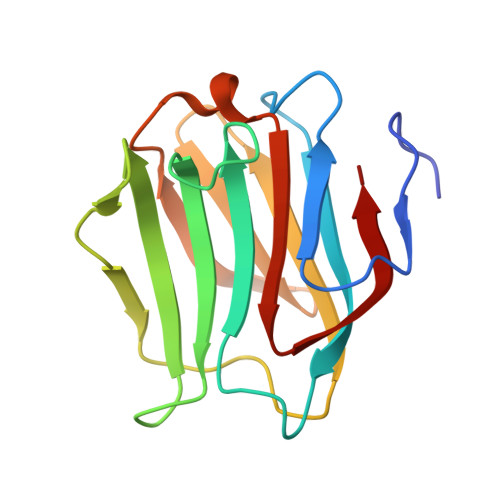
Crystal structure of human galectin-9 N-terminal CRD in complex with N-acetyllactosamine trimer (crystal 2)


Experimental Data Snapshot

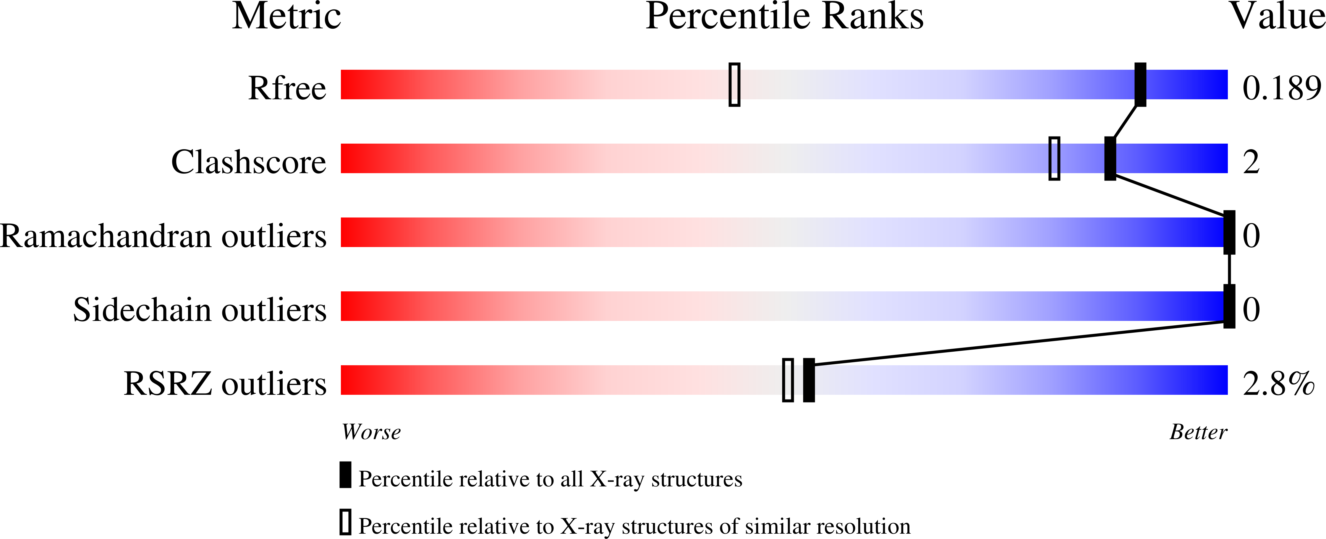
  • Method: X-RAY DIFFRACTION
  • Resolution: 1.30 Å
  • R-Value Free: 0.192 
  • R-Value Work: 0.182 
  • R-Value Observed: 0.182 

wwPDB Validation   3D Report Full Report


Ligand Structure Quality Assessment 


This is version 2.1 of the entry. See complete history


Literature

Structural analysis of the recognition mechanism of poly-N-acetyllactosamine by the human galectin-9 N-terminal carbohydrate recognition domain.

Nagae, M.Nishi, N.Murata, T.Usui, T.Nakamura, T.Wakatsuki, S.Kato, R.

(2009) Glycobiology 19: 112-117

  • DOI: https://doi.org/10.1093/glycob/cwn121
  • Primary Citation of Related Structures:  
    2ZHK, 2ZHL, 2ZHM, 2ZHN

  • PubMed Abstract: 

    Galectins are a family of beta-galactoside-specific lectins bearing a conserved carbohydrate recognition domain. Interactions between galectins and poly-N-acetyllactosamine sequences are critical in a variety of biological processes. Galectin-9, a member of the galectin family, has two carbohydrate recognition domains at both the N- and C-terminal regions. Here we report the crystal structure of the human galectin-9 N-terminal carbohydrate recognition domain in complex with N-acetyllactosamine dimers and trimers. These complex structures revealed that the galectin-9 N-terminal carbohydrate recognition domain can recognize internal N-acetyllactosamine units within poly-N-acetyllactosamine chains. Based on these complex structures, we propose two putative recognition modes for poly-N-acetyllactosamine binding by galectins.


  • Organizational Affiliation

    Structural Biology Research Center, Photon Factory, Institute of Materials Structure Science, High Energy Accelerator Research Organization (KEK), Tsukuba, Ibaraki, Japan.


Macromolecules
Find similar proteins by:  (by identity cutoff)  |  3D Structure
Entity ID: 1
MoleculeChains Sequence LengthOrganismDetailsImage
GALECTIN-9148Homo sapiensMutation(s): 0 
UniProt & NIH Common Fund Data Resources
Find proteins for O00182 (Homo sapiens)
Explore O00182 
Go to UniProtKB:  O00182
PHAROS:  O00182
GTEx:  ENSG00000168961 
Entity Groups  
Sequence Clusters30% Identity50% Identity70% Identity90% Identity95% Identity100% Identity
UniProt GroupO00182
Sequence Annotations
Expand
  • Reference Sequence
Oligosaccharides

Help

Entity ID: 2
MoleculeChains Length2D Diagram Glycosylation3D Interactions
beta-D-galactopyranose-(1-4)-2-acetamido-2-deoxy-beta-D-glucopyranose-(1-3)-beta-D-galactopyranose-(1-4)-2-acetamido-2-deoxy-beta-D-glucopyranose-(1-3)-beta-D-galactopyranose-(1-4)-2-acetamido-2-deoxy-beta-D-glucopyranose
B
6N/A
Glycosylation Resources
GlyTouCan:  G46055MA
GlyCosmos:  G46055MA
GlyGen:  G46055MA
Small Molecules
Ligands 1 Unique
IDChains Name / Formula / InChI Key2D Diagram3D Interactions
RPN
Query on RPN

Download Ideal Coordinates CCD File 
C [auth A](R)-1-PARA-NITRO-PHENYL-2-AZIDO-ETHANOL
C8 H8 N4 O3
DHEGJYKMZJGYGW-QMMMGPOBSA-N
Experimental Data & Validation

Experimental Data

  • Method: X-RAY DIFFRACTION
  • Resolution: 1.30 Å
  • R-Value Free: 0.192 
  • R-Value Work: 0.182 
  • R-Value Observed: 0.182 
  • Space Group: P 41 21 2
Unit Cell:
Length ( Å )Angle ( ˚ )
a = 52.157α = 90
b = 52.157β = 90
c = 113.83γ = 90
Software Package:
Software NamePurpose
MOLREPphasing
REFMACrefinement
HKL-2000data reduction
HKL-2000data scaling

Structure Validation

View Full Validation Report


Ligand Structure Quality Assessment 


Entry History 

Deposition Data

Revision History  (Full details and data files)

  • Version 1.0: 2008-12-30
    Type: Initial release
  • Version 1.1: 2011-07-13
    Changes: Non-polymer description, Version format compliance
  • Version 2.0: 2020-07-29
    Type: Remediation
    Reason: Carbohydrate remediation
    Changes: Advisory, Atomic model, Data collection, Derived calculations, Structure summary
  • Version 2.1: 2024-03-13
    Changes: Data collection, Database references, Structure summary