2YCL

complete structure of the corrinoid,iron-sulfur protein including the N-terminal domain with a 4Fe-4S cluster


Experimental Data Snapshot

  • Method: X-RAY DIFFRACTION
  • Resolution: 1.95 Å
  • R-Value Free: 0.251 
  • R-Value Work: 0.192 
  • R-Value Observed: 0.195 

Starting Model: experimental
View more details

wwPDB Validation   3D Report Full Report


Ligand Structure Quality Assessment 


This is version 1.3 of the entry. See complete history


Literature

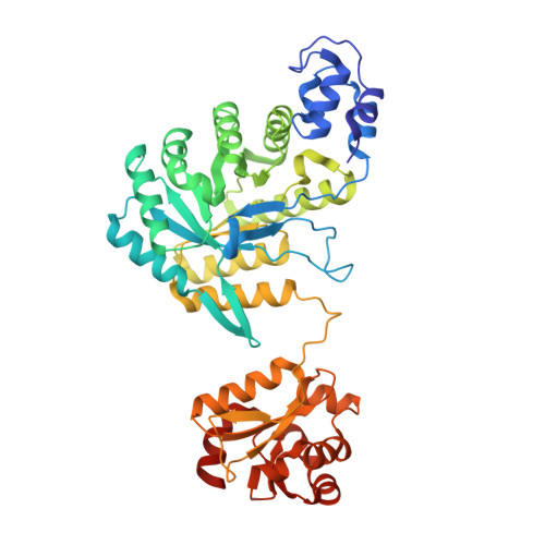
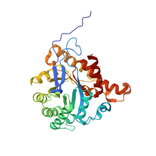
Structural Basis for Electron and Methyl-Group Transfer in a Methyltransferase System Operating in the Reductive Acetyl-Coa Pathway

Goetzl, S.Jeoung, J.H.Hennig, S.E.Dobbek, H.

(2011) J Mol Biol 411: 96

  • DOI: https://doi.org/10.1016/j.jmb.2011.05.025
  • Primary Citation of Related Structures:  
    2YCI, 2YCJ, 2YCK, 2YCL

  • PubMed Abstract: 

    Several anaerobic acetogenic, methanogenic, hydrogenogenic, and sulfate-reducing microorganisms are able to use the reductive acetyl-CoA (Wood-Ljungdahl) pathway to convert CO₂ into biomass. The reductive acetyl-CoA pathway consists of two branches connected by the Co/Fe-containing corrinoid iron-sulfur protein (CoFeSP), which transfers a methyl group from a methyltransferase (MeTr)/methyltetrahydrofolate (CH₃-H₄ folate) complex to the reduced Ni-Ni-[4Fe-4S] cluster (cluster A) of acetyl-CoA synthase. We investigated the CoFeSP and MeTr couple of the hydrogenogenic bacterium Carboxydothermus hydrogenoformans and show that the two proteins are able to catalyze the methyl-group transfer reaction from CH₃-H₄ folate to the Co(I) center of CoFeSP. We determined the crystal structures of both proteins. The structure of CoFeSP includes the previously unresolved N-terminal domain of the large subunit of CoFeSP, revealing a unique four-helix-bundle-like architecture in which a [4Fe-4S] cluster is shielded by hydrophobic amino acids. It further reveals that the corrinoid and the [4Fe-4S] cluster binding domains are mobile, which is mandatory for the postulated electron transfer between them. Furthermore, we solved the crystal structures of apo-MeTr, CH₃-H₄-folate-bound MeTr, and H₄-folate-bound MeTr, revealing a substrate-induced closure of the CH₃-H₄ folate binding cavity of MeTr. We observed three different conformations of Asn200 depending on the substrate bound in the active site, demonstrating its conformational modulation by hydrogen-bonding interactions with the substrate. The observed flexibility could be essential to stabilize the transition state during methyl-group transfer. The conformational space and role of Asn200 are likely conserved in homologous cobalamin-dependent MeTrs such as methionine synthase.


  • Organizational Affiliation

    Institut für Biologie, Strukturbiologie/Biochemie, Humboldt-Universität zu Berlin, Leonor-Michaelis-Haus, 10115 Berlin, Germany.


Macromolecules
Find similar proteins by:  (by identity cutoff)  |  3D Structure
Entity ID: 1
MoleculeChains Sequence LengthOrganismDetailsImage
CARBON MONOXIDE DEHYDROGENASE CORRINOID/IRON-SULFUR PROTEIN, GAMMA SUBUNIT445Carboxydothermus hydrogenoformans Z-2901Mutation(s): 0 
UniProt
Find proteins for Q3ACS3 (Carboxydothermus hydrogenoformans (strain ATCC BAA-161 / DSM 6008 / Z-2901))
Explore Q3ACS3 
Go to UniProtKB:  Q3ACS3
Entity Groups  
Sequence Clusters30% Identity50% Identity70% Identity90% Identity95% Identity100% Identity
UniProt GroupQ3ACS3
Sequence Annotations
Expand
  • Reference Sequence
Find similar proteins by:  (by identity cutoff)  |  3D Structure
Entity ID: 2
MoleculeChains Sequence LengthOrganismDetailsImage
CO DEHYDROGENASE/ACETYL-COA SYNTHASE, IRON-SULFUR PROTEIN310Carboxydothermus hydrogenoformans Z-2901Mutation(s): 0 
UniProt
Find proteins for Q3ACS0 (Carboxydothermus hydrogenoformans (strain ATCC BAA-161 / DSM 6008 / Z-2901))
Explore Q3ACS0 
Go to UniProtKB:  Q3ACS0
Entity Groups  
Sequence Clusters30% Identity50% Identity70% Identity90% Identity95% Identity100% Identity
UniProt GroupQ3ACS0
Sequence Annotations
Expand
  • Reference Sequence
Small Molecules
Ligands 2 Unique
IDChains Name / Formula / InChI Key2D Diagram3D Interactions
B12
Query on B12

Download Ideal Coordinates CCD File 
C [auth A]COBALAMIN
C62 H89 Co N13 O14 P
LKVIQTCSMMVGFU-DWSMJLPVSA-N
SF4
Query on SF4

Download Ideal Coordinates CCD File 
D [auth A]IRON/SULFUR CLUSTER
Fe4 S4
LJBDFODJNLIPKO-UHFFFAOYSA-N
Experimental Data & Validation

Experimental Data

  • Method: X-RAY DIFFRACTION
  • Resolution: 1.95 Å
  • R-Value Free: 0.251 
  • R-Value Work: 0.192 
  • R-Value Observed: 0.195 
  • Space Group: C 1 2 1
Unit Cell:
Length ( Å )Angle ( ˚ )
a = 172.219α = 90
b = 43.476β = 119.39
c = 101.855γ = 90
Software Package:
Software NamePurpose
PHENIXrefinement
PHASERphasing

Structure Validation

View Full Validation Report


Ligand Structure Quality Assessment 


Entry History 

Deposition Data

Revision History  (Full details and data files)

  • Version 1.0: 2011-06-08
    Type: Initial release
  • Version 1.1: 2011-08-03
    Changes: Database references, Other, Version format compliance
  • Version 1.2: 2012-10-24
    Changes: Non-polymer description
  • Version 1.3: 2023-12-20
    Changes: Data collection, Database references, Derived calculations, Other, Refinement description