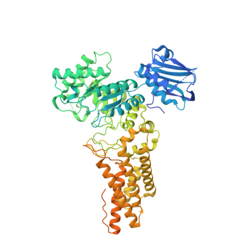
2WZH

BtGH84 D242N in complex with MeUMB-derived oxazoline


Experimental Data Snapshot

  • Method: X-RAY DIFFRACTION
  • Resolution: 2.20 Å
  • R-Value Free: 0.227 
  • R-Value Work: 0.186 
  • R-Value Observed: 0.188 

Starting Model: experimental
View more details

wwPDB Validation   3D Report Full Report


Ligand Structure Quality Assessment 


This is version 1.2 of the entry. See complete history


Literature

Visualizing the Reaction Coordinate of an O-Glcnac Hydrolase

He, Y.Macauley, M.S.Stubbs, K.A.Vocadlo, D.J.Davies, G.J.

(2010) J Am Chem Soc 132: 1807

  • DOI: https://doi.org/10.1021/ja9086769
  • Primary Citation of Related Structures:  
    2WZH, 2WZI, 2X0H

  • PubMed Abstract: 

    N-Acetylglucosamine beta-O-linked to serine and threonine residues of nucleocytoplasmic proteins (O-GlcNAc) has been linked to neurodegeneration, cellular stress response, and transcriptional regulation. Removal of O-GlcNAc is catalyzed by O-GlcNAcase (OGA) using a substrate-assisted catalytic mechanism. Here we define the reaction coordinate using chemical approaches and directly observe both a Michaelis complex and the oxazoline intermediate.


  • Organizational Affiliation

    York Structural Biology laboratory, Department of Chemistry, The University of York, YO10 5YW, UK.


Macromolecules
Find similar proteins by:  (by identity cutoff)  |  3D Structure
Entity ID: 1
MoleculeChains Sequence LengthOrganismDetailsImage
O-GLCNACASE BT_4395737Bacteroides thetaiotaomicron VPI-5482Mutation(s): 1 
EC: 3.2.1.52
UniProt
Find proteins for Q89ZI2 (Bacteroides thetaiotaomicron (strain ATCC 29148 / DSM 2079 / JCM 5827 / CCUG 10774 / NCTC 10582 / VPI-5482 / E50))
Explore Q89ZI2 
Go to UniProtKB:  Q89ZI2
Entity Groups  
Sequence Clusters30% Identity50% Identity70% Identity90% Identity95% Identity100% Identity
UniProt GroupQ89ZI2
Sequence Annotations
Expand
  • Reference Sequence
Small Molecules
Ligands 3 Unique
IDChains Name / Formula / InChI Key2D Diagram3D Interactions
NGO
Query on NGO

Download Ideal Coordinates CCD File 
B [auth A]2-METHYL-4,5-DIHYDRO-(1,2-DIDEOXY-ALPHA-D-GLUCOPYRANOSO)[2,1-D]-1,3-OXAZOLE
C8 H14 N O5
PDBSWNMXMILYCQ-PVFLNQBWSA-O
GOL
Query on GOL

Download Ideal Coordinates CCD File 
C [auth A]GLYCEROL
C3 H8 O3
PEDCQBHIVMGVHV-UHFFFAOYSA-N
CA
Query on CA

Download Ideal Coordinates CCD File 
D [auth A]CALCIUM ION
Ca
BHPQYMZQTOCNFJ-UHFFFAOYSA-N
Experimental Data & Validation

Experimental Data

  • Method: X-RAY DIFFRACTION
  • Resolution: 2.20 Å
  • R-Value Free: 0.227 
  • R-Value Work: 0.186 
  • R-Value Observed: 0.188 
  • Space Group: C 1 2 1
Unit Cell:
Length ( Å )Angle ( ˚ )
a = 184.76α = 90
b = 52.29β = 100.2
c = 86.93γ = 90
Software Package:
Software NamePurpose
REFMACrefinement
MOSFLMdata reduction
SCALAdata scaling
PHASERphasing

Structure Validation

View Full Validation Report


Ligand Structure Quality Assessment 


Entry History 

Deposition Data

Revision History  (Full details and data files)

  • Version 1.0: 2010-01-26
    Type: Initial release
  • Version 1.1: 2011-10-26
    Changes: Database references, Derived calculations, Non-polymer description, Refinement description, Version format compliance
  • Version 1.2: 2023-12-20
    Changes: Data collection, Database references, Derived calculations, Other, Refinement description