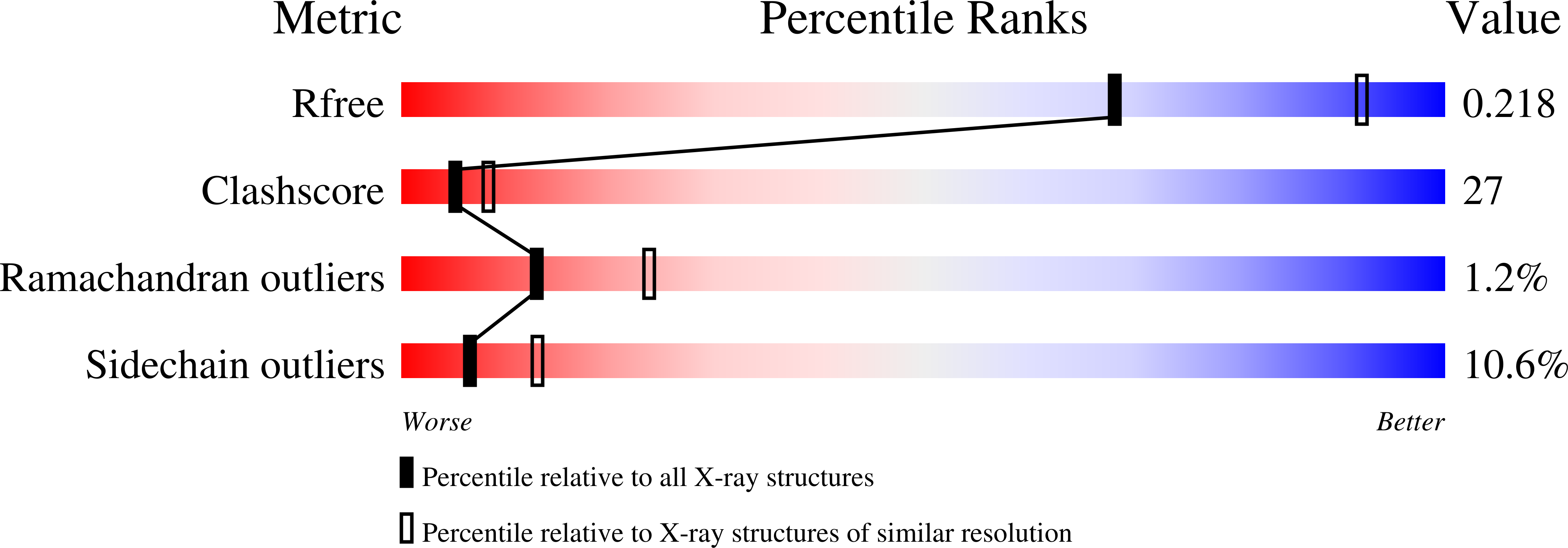
2R8V

Native structure of N-acetylglutamate synthase from Neisseria gonorrhoeae


Experimental Data Snapshot

  • Method: X-RAY DIFFRACTION
  • Resolution: 2.50 Å
  • R-Value Free: 0.252 
  • R-Value Work: 0.192 
  • R-Value Observed: 0.194 

wwPDB Validation   3D Report Full Report


This is version 1.4 of the entry. See complete history


Literature

The crystal structure of N-acetyl-L-glutamate synthase from Neisseria gonorrhoeae provides insights into mechanisms of catalysis and regulation.

Shi, D.Sagar, V.Jin, Z.Yu, X.Caldovic, L.Morizono, H.Allewell, N.M.Tuchman, M.

(2008) J Biol Chem 283: 7176-7184

  • DOI: https://doi.org/10.1074/jbc.M707678200
  • Primary Citation of Related Structures:  
    2R8V, 2R98, 3B8G

  • PubMed Abstract: 

    The crystal structures of N-acetylglutamate synthase (NAGS) in the arginine biosynthetic pathway of Neisseria gonorrhoeae complexed with acetyl-CoA and with CoA plus N-acetylglutamate have been determined at 2.5- and 2.6-A resolution, respectively. The monomer consists of two separately folded domains, an amino acid kinase (AAK) domain and an N-acetyltransferase (NAT) domain connected through a 10-A linker. The monomers assemble into a hexameric ring that consists of a trimer of dimers with 32-point symmetry, inner and outer ring diameters of 20 and 100A, respectively, and a height of 110A(.) Each AAK domain interacts with the cognate domains of two adjacent monomers across two 2-fold symmetry axes and with the NAT domain from a second monomer of the adjacent dimer in the ring. The catalytic sites are located within the NAT domains. Three active site residues, Arg316, Arg425, and Ser427, anchor N-acetylglutamate in a position at the active site to form hydrogen bond interactions to the main chain nitrogen atoms of Cys356 and Leu314, and hydrophobic interactions to the side chains of Leu313 and Leu314. The mode of binding of acetyl-CoA and CoA is similar to other NAT family proteins. The AAK domain, although catalytically inactive, appears to bind arginine. This is the first reported crystal structure of any NAGS, and it provides insights into the catalytic function and arginine regulation of NAGS enzymes.


  • Organizational Affiliation

    Children's Research Institute, Children's National Medical Center, The George Washington University, Washington, DC 20010, USA. dshi@cnmcresearch.org


Macromolecules
Find similar proteins by:  (by identity cutoff)  |  3D Structure
Entity ID: 1
MoleculeChains Sequence LengthOrganismDetailsImage
Putative acetylglutamate synthase456Neisseria gonorrhoeaeMutation(s): 3 
Gene Names: argA
EC: 2.3.1.1
UniProt
Find proteins for Q5FAK7 (Neisseria gonorrhoeae (strain ATCC 700825 / FA 1090))
Explore Q5FAK7 
Go to UniProtKB:  Q5FAK7
Entity Groups  
Sequence Clusters30% Identity50% Identity70% Identity90% Identity95% Identity100% Identity
UniProt GroupQ5FAK7
Sequence Annotations
Expand
  • Reference Sequence
Small Molecules
Ligands 1 Unique
IDChains Name / Formula / InChI Key2D Diagram3D Interactions
ACO
Query on ACO

Download Ideal Coordinates CCD File 
B [auth A]ACETYL COENZYME *A
C23 H38 N7 O17 P3 S
ZSLZBFCDCINBPY-ZSJPKINUSA-N
Experimental Data & Validation

Experimental Data

  • Method: X-RAY DIFFRACTION
  • Resolution: 2.50 Å
  • R-Value Free: 0.252 
  • R-Value Work: 0.192 
  • R-Value Observed: 0.194 
  • Space Group: P 3 1 2
Unit Cell:
Length ( Å )Angle ( ˚ )
a = 98.661α = 90
b = 98.661β = 90
c = 89.755γ = 120
Software Package:
Software NamePurpose
REFMACrefinement
CrystalCleardata collection
HKL-2000data reduction
HKL-2000data scaling
SHELXSphasing
CNSrefinement

Structure Validation

View Full Validation Report


Entry History 

Deposition Data

Revision History  (Full details and data files)

  • Version 1.0: 2008-01-15
    Type: Initial release
  • Version 1.1: 2011-07-13
    Changes: Advisory, Version format compliance
  • Version 1.2: 2019-07-24
    Changes: Data collection, Refinement description
  • Version 1.3: 2021-10-20
    Changes: Database references, Derived calculations
  • Version 1.4: 2024-02-21
    Changes: Data collection