2KIN

KINESIN (MONOMERIC) FROM RATTUS NORVEGICUS


Experimental Data Snapshot

  • Method: X-RAY DIFFRACTION
  • Resolution: 2.00 Å
  • R-Value Free: 0.254 
  • R-Value Work: 0.194 

wwPDB Validation   3D Report Full Report


This is version 1.4 of the entry. See complete history


Literature

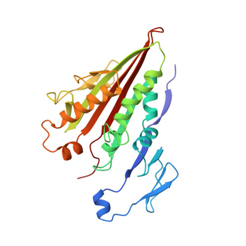
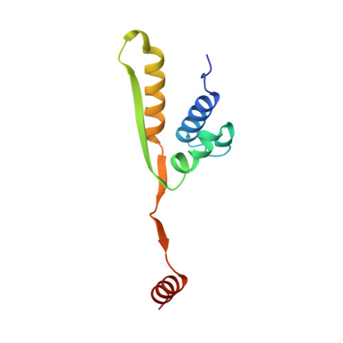
X-ray structure of motor and neck domains from rat brain kinesin.

Sack, S.Muller, J.Marx, A.Thormahlen, M.Mandelkow, E.M.Brady, S.T.Mandelkow, E.

(1997) Biochemistry 36: 16155-16165

  • DOI: https://doi.org/10.1021/bi9722498
  • Primary Citation of Related Structures:  
    2KIN

  • PubMed Abstract: 

    We have determined the X-ray structure of rat kinesin head and neck domains. The folding of the core motor domain resembles that of human kinesin reported recently [Kull, F. J., et al. (1996) Nature 380, 550-554]. Novel features of the structure include the N-terminal region, folded as a beta-strand, and the C-terminal transition from the motor to the rod domain, folded as two beta-strands plus an alpha-helix. This helix is the beginning of kinesin's neck responsible for dimerization of the motor complex and for force transduction. Although the folding of the motor domain core is similar to that of a domain of myosin (an actin-dependent motor), the position and angle of kinesin's neck are very different from those of myosin's stalk, suggesting that the two motors have different mechanisms of force transduction. The N- and C-terminal ends of the core motor, thought to be responsible for the directionality of the motors [Case, R. B., et al. (1997) Cell 90, 959-966], take the form of beta-strands attached to the central beta-sheet of the structure.


  • Organizational Affiliation

    Max-Planck-Unit for Structural Molecular Biology, c/o DESY, Notkestrasse 85, D-22607 Hamburg, Germany.


Macromolecules
Find similar proteins by:  (by identity cutoff)  |  3D Structure
Entity ID: 1
MoleculeChains Sequence LengthOrganismDetailsImage
KINESIN238Rattus norvegicusMutation(s): 0 
UniProt
Find proteins for P56536 (Rattus norvegicus)
Explore P56536 
Go to UniProtKB:  P56536
Entity Groups  
Sequence Clusters30% Identity50% Identity70% Identity90% Identity95% Identity100% Identity
UniProt GroupP56536
Sequence Annotations
Expand
  • Reference Sequence
Find similar proteins by:  (by identity cutoff)  |  3D Structure
Entity ID: 2
MoleculeChains Sequence LengthOrganismDetailsImage
KINESIN100Rattus norvegicusMutation(s): 0 
UniProt
Find proteins for Q6QLM7 (Rattus norvegicus)
Explore Q6QLM7 
Go to UniProtKB:  Q6QLM7
Entity Groups  
Sequence Clusters30% Identity50% Identity70% Identity90% Identity95% Identity100% Identity
UniProt GroupQ6QLM7
Sequence Annotations
Expand
  • Reference Sequence
Small Molecules
Experimental Data & Validation

Experimental Data

  • Method: X-RAY DIFFRACTION
  • Resolution: 2.00 Å
  • R-Value Free: 0.254 
  • R-Value Work: 0.194 
  • Space Group: P 21 21 21
Unit Cell:
Length ( Å )Angle ( ˚ )
a = 71.556α = 90
b = 73.674β = 90
c = 74.128γ = 90
Software Package:
Software NamePurpose
AMoREphasing
REFMACrefinement
DENZOdata reduction
SCALEPACKdata scaling

Structure Validation

View Full Validation Report


Entry History 

Deposition Data

Revision History  (Full details and data files)

  • Version 1.0: 1998-04-08
    Type: Initial release
  • Version 1.1: 2008-03-24
    Changes: Version format compliance
  • Version 1.2: 2011-07-13
    Changes: Version format compliance
  • Version 1.3: 2024-02-21
    Changes: Data collection, Database references, Derived calculations
  • Version 1.4: 2024-04-03
    Changes: Refinement description