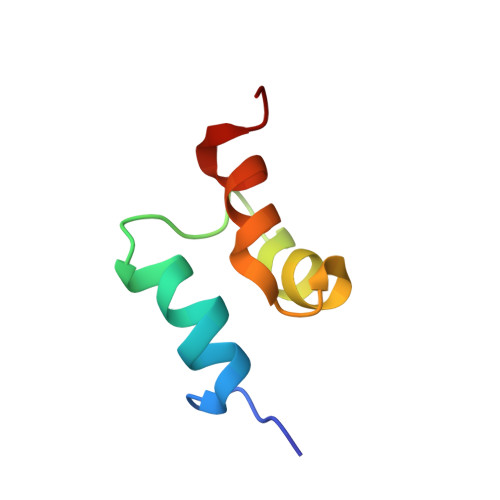
Ubiquitin Recognition by the Ubiquitin-associated Domain of p62 Involves a Novel Conformational Switch
Long, J., Gallagher, T.R., Cavey, J.R., Sheppard, P.W., Ralston, S.H., Layfield, R., Searle, M.S.(2008) J Biol Chem 283: 5427-5440
- PubMed: 18083707
- DOI: https://doi.org/10.1074/jbc.M704973200
- Primary Citation of Related Structures:
2JY7, 2JY8 - PubMed Abstract:
The p62 protein functions as a scaffold in signaling pathways that lead to activation of NF-kappaB and is an important regulator of osteoclastogenesis. Mutations affecting the receptor activator of NF-kappaB signaling axis can result in human skeletal disorders, including those identified in the C-terminal ubiquitin-associated (UBA) domain of p62 in patients with Paget disease of bone. These observations suggest that the disease may involve a common mechanism related to alterations in the ubiquitin-binding properties of p62. The structural basis for ubiquitin recognition by the UBA domain of p62 has been investigated using NMR and reveals a novel binding mechanism involving a slow exchange structural reorganization of the UBA domain to a "bound" non-canonical UBA conformation that is not significantly populated in the absence of ubiquitin. The repacking of the three-helix bundle generates a binding surface localized around the conserved Xaa-Gly-Phe-Xaa loop that appears to optimize both hydrophobic and electrostatic surface complementarity with ubiquitin. NMR titration analysis shows that the p62-UBA binds to Lys 48-linked di-ubiquitin with approximately 4-fold lower affinity than to mono-ubiquitin, suggesting preferential binding of the p62-UBA to single ubiquitin units, consistent with the apparent in vivo preference of the p62 protein for Lys 63-linked polyubiquitin chains (which adopt a more open and extended structure). The conformational switch observed on binding may represent a novel mechanism that underlies specificity in regulating signalinduced protein recognition events.
Organizational Affiliation:
School of Chemistry, Centre for Biomolecular Sciences, University of Nottingham, Nottingham NG7 2RD, United Kingdom.


