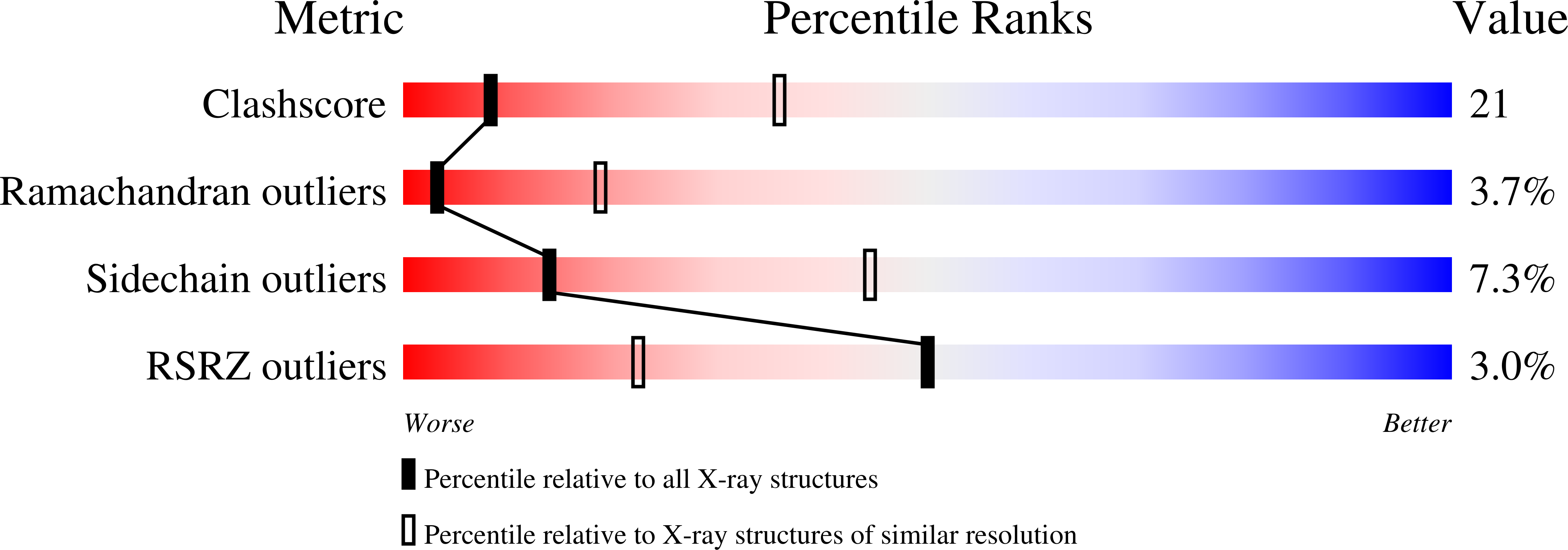
2IHN

Co-crystal of Bacteriophage T4 RNase H with a fork DNA substrate


Experimental Data Snapshot

  • Method: X-RAY DIFFRACTION
  • Resolution: 3.00 Å
  • R-Value Free: 0.282 
  • R-Value Work: 0.222 
  • R-Value Observed: 0.225 

Starting Model: experimental
View more details

wwPDB Validation   3D Report Full Report


This is version 1.3 of the entry. See complete history


Literature

Crystal structure of bacteriophage T4 5' nuclease in complex with a branched DNA reveals how FEN-1 family nucleases bind their substrates.

Devos, J.M.Tomanicek, S.J.Jones, C.E.Nossal, N.G.Mueser, T.C.

(2007) J Biol Chem 282: 31713-31724

  • DOI: https://doi.org/10.1074/jbc.M703209200
  • Primary Citation of Related Structures:  
    2IHN

  • PubMed Abstract: 

    Bacteriophage T4 RNase H, a flap endonuclease-1 family nuclease, removes RNA primers from lagging strand fragments. It has both 5' nuclease and flap endonuclease activities. Our previous structure of native T4 RNase H (PDB code 1TFR) revealed an active site composed of highly conserved Asp residues and two bound hydrated magnesium ions. Here, we report the crystal structure of T4 RNase H in complex with a fork DNA substrate bound in its active site. This is the first structure of a flap endonuclease-1 family protein with its complete branched substrate. The fork duplex interacts with an extended loop of the helix-hairpin-helix motif class 2. The 5' arm crosses over the active site, extending below the bridge (helical arch) region. Cleavage assays of this DNA substrate identify a primary cut site 7-bases in from the 5' arm. The scissile phosphate, the first bond in the duplex DNA adjacent to the 5' arm, lies above a magnesium binding site. The less ordered 3' arm reaches toward the C and N termini of the enzyme, which are binding sites for T4 32 protein and T4 45 clamp, respectively. In the crystal structure, the scissile bond is located within the double-stranded DNA, between the first two duplex nucleotides next to the 5' arm, and lies above a magnesium binding site. This complex provides important insight into substrate recognition and specificity of the flap endonuclease-1 enzymes.


  • Organizational Affiliation

    Department of Chemistry, The University of Toledo, Toledo, Ohio 43606, USA.


Macromolecules

Find similar proteins by:  (by identity cutoff)  |  3D Structure
Entity ID: 3
MoleculeChains Sequence LengthOrganismDetailsImage
Ribonuclease HC [auth A]305Tequatrovirus T4Mutation(s): 1 
Gene Names: rnh33.2das
EC: 3.1.26.4
UniProt
Find proteins for P13319 (Enterobacteria phage T4)
Explore P13319 
Go to UniProtKB:  P13319
Entity Groups  
Sequence Clusters30% Identity50% Identity70% Identity90% Identity95% Identity100% Identity
UniProt GroupP13319
Sequence Annotations
Expand
  • Reference Sequence

Find similar nucleic acids by:  Sequence   |   3D Structure  

Entity ID: 1
MoleculeChains LengthOrganismImage
5'-D(*CP*TP*AP*AP*CP*TP*TP*TP*GP*AP*GP*GP*CP*AP*GP*AP*CP*C)-3'A [auth C]18N/A
Sequence Annotations
Expand
  • Reference Sequence

Find similar nucleic acids by:  Sequence   |   3D Structure  

Entity ID: 2
MoleculeChains LengthOrganismImage
5'-D(*GP*GP*TP*CP*TP*GP*CP*CP*TP*CP*AP*AP*GP*AP*CP*GP*GP*TP*AP*GP*TP*CP*AP*A)-3'B [auth D]24N/A
Sequence Annotations
Expand
  • Reference Sequence
Experimental Data & Validation

Experimental Data

  • Method: X-RAY DIFFRACTION
  • Resolution: 3.00 Å
  • R-Value Free: 0.282 
  • R-Value Work: 0.222 
  • R-Value Observed: 0.225 
  • Space Group: P 21 21 21
Unit Cell:
Length ( Å )Angle ( ˚ )
a = 68.16α = 90
b = 84.99β = 90
c = 89.29γ = 90
Software Package:
Software NamePurpose
REFMACrefinement
CrystalCleardata reduction
AMoREphasing

Structure Validation

View Full Validation Report


Entry History 

Deposition Data

Revision History  (Full details and data files)

  • Version 1.0: 2007-08-21
    Type: Initial release
  • Version 1.1: 2011-07-13
    Changes: Version format compliance
  • Version 1.2: 2021-10-20
    Changes: Database references
  • Version 1.3: 2023-08-30
    Changes: Data collection, Refinement description