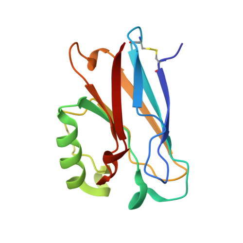
2I7S

Crystal structure of Re(phen)(CO)3 (Thr124His)(His83Gln) Azurin Cu(II) from Pseudomonas aeruginosa


Experimental Data Snapshot

  • Method: X-RAY DIFFRACTION
  • Resolution: 1.35 Å
  • R-Value Free: 0.239 
  • R-Value Work: 0.205 

Starting Model: experimental
View more details

wwPDB Validation   3D Report Full Report


Ligand Structure Quality Assessment 


This is version 1.4 of the entry. See complete history


Literature

Relaxation dynamics of Pseudomonas aeruginosa Re(I)(CO)3(alpha-diimine)(HisX)+ (X = 83, 107, 109, 124, 126)Cu(II) azurins.

Blanco-Rodriguez, A.M.Busby, M.Ronayne, K.Towrie, M.Gradinaru, C.Sudhamsu, J.Sykora, J.Hof, M.Zalis, S.Di Bilio, A.J.Crane, B.R.Gray, H.B.Vlcek, A.

(2009) J Am Chem Soc 131: 11788-11800

  • DOI: https://doi.org/10.1021/ja902744s
  • Primary Citation of Related Structures:  
    2I7S, 3IBO

  • PubMed Abstract: 

    Photoinduced relaxation processes of five structurally characterized Pseudomonas aeruginosa Re(I)(CO)(3)(alpha-diimine)(HisX) (X = 83, 107, 109, 124, 126)Cu(II) azurins have been investigated by time-resolved (ps-ns) IR spectroscopy and emission spectroscopy. Crystal structures reveal the presence of Re-azurin dimers and trimers that in two cases (X = 107, 124) involve van der Waals interactions between interdigitated diimine aromatic rings. Time-dependent emission anisotropy measurements confirm that the proteins aggregate in mM solutions (D(2)O, KP(i) buffer, pD = 7.1). Excited-state DFT calculations show that extensive charge redistribution in the Re(I)(CO)(3) --> diimine (3)MLCT state occurs: excitation of this (3)MLCT state triggers several relaxation processes in Re-azurins whose kinetics strongly depend on the location of the metallolabel on the protein surface. Relaxation is manifested by dynamic blue shifts of excited-state nu(CO) IR bands that occur with triexponential kinetics: intramolecular vibrational redistribution together with vibrational and solvent relaxation give rise to subps, approximately 2, and 8-20 ps components, while the approximately 10(2) ps kinetics are attributed to displacement (reorientation) of the Re(I)(CO)(3)(phen)(im) unit relative to the peptide chain, which optimizes Coulombic interactions of the Re(I) excited-state electron density with solvated peptide groups. Evidence also suggests that additional segmental movements of Re-bearing beta-strands occur without perturbing the reaction field or interactions with the peptide. Our work demonstrates that time-resolved IR spectroscopy and emission anisotropy of Re(I) carbonyl-diimine complexes are powerful probes of molecular dynamics at or around the surfaces of proteins and protein-protein interfacial regions.


  • Organizational Affiliation

    School of Biological and Chemical Sciences, Queen Mary, University of London, Mile End Road, London E1 4NS, United Kingdom.


Macromolecules
Find similar proteins by:  (by identity cutoff)  |  3D Structure
Entity ID: 1
MoleculeChains Sequence LengthOrganismDetailsImage
Azurin
A, B, C, D
128Pseudomonas aeruginosaMutation(s): 2 
Gene Names: azu
UniProt
Find proteins for P00282 (Pseudomonas aeruginosa (strain ATCC 15692 / DSM 22644 / CIP 104116 / JCM 14847 / LMG 12228 / 1C / PRS 101 / PAO1))
Explore P00282 
Go to UniProtKB:  P00282
Entity Groups  
Sequence Clusters30% Identity50% Identity70% Identity90% Identity95% Identity100% Identity
UniProt GroupP00282
Sequence Annotations
Expand
  • Reference Sequence
Small Molecules
Ligands 3 Unique
IDChains Name / Formula / InChI Key2D Diagram3D Interactions
REP
Query on REP

Download Ideal Coordinates CCD File 
F [auth A],
H [auth B],
K [auth C],
M [auth D]
(1,10 PHENANTHROLINE)-(TRI-CARBON MONOXIDE) RHENIUM (I)
C15 H8 N2 O3 Re
HRZLGVADJDWBIJ-UHFFFAOYSA-N
CON
Query on CON

Download Ideal Coordinates CCD File 
I [auth B]COBALT TETRAAMMINE ION
Co H12 N4
AWWXFUYNCLHXPM-UHFFFAOYSA-N
CU
Query on CU

Download Ideal Coordinates CCD File 
E [auth A],
G [auth B],
J [auth C],
L [auth D]
COPPER (II) ION
Cu
JPVYNHNXODAKFH-UHFFFAOYSA-N
Experimental Data & Validation

Experimental Data

  • Method: X-RAY DIFFRACTION
  • Resolution: 1.35 Å
  • R-Value Free: 0.239 
  • R-Value Work: 0.205 
  • Space Group: P 1
Unit Cell:
Length ( Å )Angle ( ˚ )
a = 33.135α = 82.92
b = 58.98β = 78.11
c = 66.107γ = 76.68
Software Package:
Software NamePurpose
SHELXrefinement
PDB_EXTRACTdata extraction
ADSCdata collection
DENZOdata reduction
SCALEPACKdata scaling
EPMRphasing
SHELXL-97refinement

Structure Validation

View Full Validation Report


Ligand Structure Quality Assessment 


Entry History 

Deposition Data

Revision History  (Full details and data files)

  • Version 1.0: 2006-12-05
    Type: Initial release
  • Version 1.1: 2008-05-01
    Changes: Version format compliance
  • Version 1.2: 2011-07-13
    Changes: Version format compliance
  • Version 1.3: 2021-10-20
    Changes: Database references, Derived calculations
  • Version 1.4: 2023-08-30
    Changes: Data collection, Refinement description