2I3I

Structure of an ML-IAP/XIAP chimera bound to a peptidomimetic


Experimental Data Snapshot

  • Method: X-RAY DIFFRACTION
  • Resolution: 2.30 Å
  • R-Value Free: 0.224 
  • R-Value Work: 0.188 
  • R-Value Observed: 0.190 

Starting Model: experimental
View more details

wwPDB Validation   3D Report Full Report


Ligand Structure Quality Assessment 


This is version 1.5 of the entry. See complete history


Literature

Design, synthesis, and biological activity of a potent Smac mimetic that sensitizes cancer cells to apoptosis by antagonizing IAPs.

Zobel, K.Wang, L.Varfolomeev, E.Franklin, M.C.Elliott, L.O.Wallweber, H.J.Okawa, D.C.Flygare, J.A.Vucic, D.Fairbrother, W.J.Deshayes, K.

(2006) ACS Chem Biol 1: 525-533

  • DOI: https://doi.org/10.1021/cb600276q
  • Primary Citation of Related Structures:  
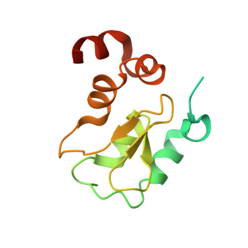
    2I3H, 2I3I

  • PubMed Abstract: 

    Designed second mitochondrial activator of caspases (Smac) mimetics based on an accessible [7,5]-bicyclic scaffold bind to and antagonize protein interactions involving the inhibitor of apoptosis (IAP) proteins, X-chromosome-linked IAP (XIAP), melanoma IAP (ML-IAP), and c-IAPs 1 and 2 (cIAP1 and cIAP2). The design rationale is based on a combination of phage-panning data, peptide binding studies, and a survey of potential isosteres. The synthesis of two scaffolds is described. These compounds bind the XIAP-baculoviral IAP repeat 3 (BIR3), cIAP1-BIR3, cIAP2-BIR3, and ML-IAP-BIR domains with submicromolar affinities. The most potent Smac mimetic binds the cIAP1-BIR3 and ML-IAP-BIR domains with a K i of 50 nM. The X-ray crystal structure of this compound bound to an ML-IAP/XIAP chimeric BIR domain protein is compared with that of a complex with a phage-derived tetrapeptide, AVPW. The structures show that these compounds bind to the Smac-binding site on ML-IAP with identical hydrogen-bonding patterns and similar hydrophobic interactions. Consistent with the structural data, coimmunoprecipitation experiments demonstrate that the compounds can effectively block Smac interactions with ML-IAP. The compounds are further demonstrated to activate caspase-3 and -7, to reduce cell viability in assays using MDA-MB-231 breast cancer cells and A2058 melanoma cells, and to enhance doxorubicin-induced apoptosis in MDA-MB-231 cells.


  • Organizational Affiliation

    Department of Protein Engineering, Genentech, Inc., South San Francisco, California 94080, USA.


Macromolecules
Find similar proteins by:  (by identity cutoff)  |  3D Structure
Entity ID: 1
MoleculeChains Sequence LengthOrganismDetailsImage
Baculoviral IAP repeat-containing protein 7
A, B
133Homo sapiensMutation(s): 0 
Gene Names: BIRC7
UniProt & NIH Common Fund Data Resources
Find proteins for Q96CA5 (Homo sapiens)
Explore Q96CA5 
Go to UniProtKB:  Q96CA5
GTEx:  ENSG00000101197 
Entity Groups  
Sequence Clusters30% Identity50% Identity70% Identity90% Identity95% Identity100% Identity
UniProt GroupQ96CA5
Sequence Annotations
Expand
  • Reference Sequence
Small Molecules
Ligands 5 Unique
IDChains Name / Formula / InChI Key2D Diagram3D Interactions
618
Query on 618

Download Ideal Coordinates CCD File 
D [auth A],
G [auth B]
(3R,6R,9AR)-2,2-DIMETHYL-6-[(N-METHYL-L-ALANYL)AMINO]-N-(3-METHYL-1-PHENYL-1H-PYRAZOL-5-YL)-5-OXO-2,3,5,6,9,9A-HEXAHYDRO[1,3]THIAZOLO[3,2-A]AZEPINE-3-CARBOXAMIDE
C25 H32 N6 O3 S
WIYCWFZPNIUIGJ-RCVZYCBYSA-N
BTB
Query on BTB

Download Ideal Coordinates CCD File 
H [auth B]2-[BIS-(2-HYDROXY-ETHYL)-AMINO]-2-HYDROXYMETHYL-PROPANE-1,3-DIOL
C8 H19 N O5
OWMVSZAMULFTJU-UHFFFAOYSA-N
ZN
Query on ZN

Download Ideal Coordinates CCD File 
C [auth A],
E [auth B]
ZINC ION
Zn
PTFCDOFLOPIGGS-UHFFFAOYSA-N
EDO
Query on EDO

Download Ideal Coordinates CCD File 
I [auth B],
J [auth B]
1,2-ETHANEDIOL
C2 H6 O2
LYCAIKOWRPUZTN-UHFFFAOYSA-N
LI
Query on LI

Download Ideal Coordinates CCD File 
F [auth B]LITHIUM ION
Li
HBBGRARXTFLTSG-UHFFFAOYSA-N
Binding Affinity Annotations 
IDSourceBinding Affinity
618 PDBBind:  2I3I Ki: 50 (nM) from 1 assay(s)
Experimental Data & Validation

Experimental Data

  • Method: X-RAY DIFFRACTION
  • Resolution: 2.30 Å
  • R-Value Free: 0.224 
  • R-Value Work: 0.188 
  • R-Value Observed: 0.190 
  • Space Group: P 41 21 2
Unit Cell:
Length ( Å )Angle ( ˚ )
a = 87.356α = 90
b = 87.356β = 90
c = 73.196γ = 90
Software Package:
Software NamePurpose
REFMACrefinement
MAR345data collection
DENZOdata reduction
SCALEPACKdata scaling

Structure Validation

View Full Validation Report


Ligand Structure Quality Assessment 


Entry History 

Deposition Data

Revision History  (Full details and data files)

  • Version 1.0: 2006-09-19
    Type: Initial release
  • Version 1.1: 2008-05-01
    Changes: Version format compliance
  • Version 1.2: 2011-07-13
    Changes: Advisory, Version format compliance
  • Version 1.3: 2017-10-18
    Changes: Refinement description
  • Version 1.4: 2024-02-21
    Changes: Data collection, Database references, Derived calculations
  • Version 1.5: 2024-04-03
    Changes: Refinement description