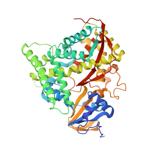
2HPD

CRYSTAL STRUCTURE OF HEMOPROTEIN DOMAIN OF P450BM-3, A PROTOTYPE FOR MICROSOMAL P450'S


Experimental Data Snapshot

  • Method: X-RAY DIFFRACTION
  • Resolution: 2.00 Å
  • R-Value Work: 0.167 
  • R-Value Observed: 0.167 

wwPDB Validation   3D Report Full Report


This is version 1.3 of the entry. See complete history


Literature

Crystal structure of hemoprotein domain of P450BM-3, a prototype for microsomal P450's.

Ravichandran, K.G.Boddupalli, S.S.Hasermann, C.A.Peterson, J.A.Deisenhofer, J.

(1993) Science 261: 731-736

  • DOI: https://doi.org/10.1126/science.8342039
  • Primary Citation of Related Structures:  
    2HPD

  • PubMed Abstract: 

    Cytochrome P450BM-3, a bacterial fatty acid monoxygenase, resembles the eukaryotic microsomal P450's and their flavoprotein reductase in primary structure and function. The three-dimensional structure of the hemoprotein domain of P450BM-3 was determined by x-ray diffraction and refined to an R factor of 16.9 percent at 2.0 angstrom resolution. The structure consists of an alph and a beta domain. The active site heme is accessible through a long hydrophobic channel formed primarily by the beta domain and the B' and F helices of the alpha domain. The two molecules in the asymmetric unit differ in conformation around the substrate binding pocket. Substantial differences between P450BM-3 and P450cam, the only other P450 structure available, are observed around the substrate binding pocket and the regions important for redox partner binding. A general mechanism for proton transfer in P450's is also proposed.


  • Organizational Affiliation

    Howard Hughes Medical Institute, University of Texas Southwestern Medical Center, Dallas 75235-9050.


Macromolecules
Find similar proteins by:  (by identity cutoff)  |  3D Structure
Entity ID: 1
MoleculeChains Sequence LengthOrganismDetailsImage
CYTOCHROME P450 BM-3
A, B
471Priestia megateriumMutation(s): 0 
EC: 1.14.14.1 (PDB Primary Data), 1.6.2.4 (UniProt)
UniProt
Find proteins for P14779 (Priestia megaterium (strain ATCC 14581 / DSM 32 / CCUG 1817 / JCM 2506 / NBRC 15308 / NCIMB 9376 / NCTC 10342 / NRRL B-14308 / VKM B-512 / Ford 19))
Explore P14779 
Go to UniProtKB:  P14779
Entity Groups  
Sequence Clusters30% Identity50% Identity70% Identity90% Identity95% Identity100% Identity
UniProt GroupP14779
Sequence Annotations
Expand
  • Reference Sequence
Small Molecules
Ligands 1 Unique
IDChains Name / Formula / InChI Key2D Diagram3D Interactions
HEM
Query on HEM

Download Ideal Coordinates CCD File 
C [auth A],
D [auth B]
PROTOPORPHYRIN IX CONTAINING FE
C34 H32 Fe N4 O4
KABFMIBPWCXCRK-RGGAHWMASA-L
Experimental Data & Validation

Experimental Data

  • Method: X-RAY DIFFRACTION
  • Resolution: 2.00 Å
  • R-Value Work: 0.167 
  • R-Value Observed: 0.167 
  • Space Group: P 1 21 1
Unit Cell:
Length ( Å )Angle ( ˚ )
a = 59.4α = 90
b = 154β = 94.7
c = 62.2γ = 90
Software Package:
Software NamePurpose
X-PLORmodel building
X-PLORrefinement
X-PLORphasing

Structure Validation

View Full Validation Report


Entry History 

Deposition Data

Revision History  (Full details and data files)

  • Version 1.0: 1993-10-31
    Type: Initial release
  • Version 1.1: 2008-03-24
    Changes: Version format compliance
  • Version 1.2: 2011-07-13
    Changes: Version format compliance
  • Version 1.3: 2024-02-14
    Changes: Data collection, Database references, Derived calculations, Other