2GYD

Complex of equine apoferritin with the H-diaziflurane photolabeling reagent


Experimental Data Snapshot

  • Method: X-RAY DIFFRACTION
  • Resolution: 1.72 Å
  • R-Value Free: 0.217 
  • R-Value Work: 0.207 
  • R-Value Observed: 0.208 

Starting Model: experimental
View more details

wwPDB Validation   3D Report Full Report


Ligand Structure Quality Assessment 


This is version 1.3 of the entry. See complete history


Literature

Photoactive analogues of the haloether anesthetics provide high-resolution features from low-affinity interactions.

Xi, J.Liu, R.Rossi, M.J.Yang, J.Loll, P.J.Dailey, W.P.Eckenhoff, R.G.

(2006) ACS Chem Biol 1: 377-384

  • DOI: https://doi.org/10.1021/cb600207d
  • Primary Citation of Related Structures:  
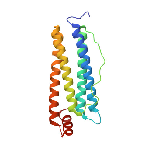
    2GYD

  • PubMed Abstract: 

    The difficulty in obtaining binding target and site information for low-affinity drugs, like the inhaled anesthetics, has limited identification of their molecular effectors. Because such information can be provided by photoactive analogues, we designed, synthesized, and characterized a novel diazirnyl haloether that closely mimics isoflurane, the most widely used clinical general anesthetic. This compound, H-diaziflurane, is a nontoxic, potent anesthetic that potentiates GABA-gated ion channels in primary cultures of hippocampal neurons. Calorimetric and structural characterizations show that H-diaziflurane binds a model anesthetic host protein with similar energetics as isoflurane and forms photoadducts with residues lining the isoflurane binding site. H-diaziflurane will be immediately useful for identifying targets and sites important for the molecular pharmacology of the inhaled haloether anesthetics.


  • Organizational Affiliation

    Department of Anesthesiology & Critical Care, University of Pennsylvania, Philadelphia, Pennsylvania 19104, USA.


Macromolecules
Find similar proteins by:  (by identity cutoff)  |  3D Structure
Entity ID: 1
MoleculeChains Sequence LengthOrganismDetailsImage
ferritin L subunit170Equus caballusMutation(s): 0 
UniProt
Find proteins for P02791 (Equus caballus)
Explore P02791 
Go to UniProtKB:  P02791
Entity Groups  
Sequence Clusters30% Identity50% Identity70% Identity90% Identity95% Identity100% Identity
UniProt GroupP02791
Sequence Annotations
Expand
  • Reference Sequence
Experimental Data & Validation

Experimental Data

  • Method: X-RAY DIFFRACTION
  • Resolution: 1.72 Å
  • R-Value Free: 0.217 
  • R-Value Work: 0.207 
  • R-Value Observed: 0.208 
  • Space Group: F 4 3 2
Unit Cell:
Length ( Å )Angle ( ˚ )
a = 181.89α = 90
b = 181.89β = 90
c = 181.89γ = 90
Software Package:
Software NamePurpose
d*TREKdata scaling
REFMACrefinement
PDB_EXTRACTdata extraction
ADSCdata collection
XTALVIEWrefinement

Structure Validation

View Full Validation Report


Ligand Structure Quality Assessment 


Entry History 

Deposition Data

Revision History  (Full details and data files)

  • Version 1.0: 2006-06-13
    Type: Initial release
  • Version 1.1: 2008-05-01
    Changes: Version format compliance
  • Version 1.2: 2011-07-13
    Changes: Derived calculations, Version format compliance
  • Version 1.3: 2023-08-30
    Changes: Data collection, Database references, Derived calculations, Refinement description