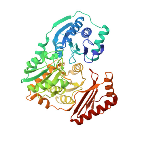
2FKM

PMM/PGM S108D mutant with alpha-d-glucose 1,6-bisphosphate bound


Experimental Data Snapshot

  • Method: X-RAY DIFFRACTION
  • Resolution: 1.90 Å
  • R-Value Free: 0.251 
  • R-Value Work: 0.203 
  • R-Value Observed: 0.205 

Starting Model: experimental
View more details

wwPDB Validation   3D Report Full Report


Ligand Structure Quality Assessment 


This is version 1.6 of the entry. See complete history


Literature

The reaction of phosphohexomutase from Pseudomonas aeruginosa: structural insights into a simple processive enzyme.

Regni, C.Schramm, A.M.Beamer, L.J.

(2006) J Biol Chem 281: 15564-15571

  • DOI: https://doi.org/10.1074/jbc.M600590200
  • Primary Citation of Related Structures:  
    2FKF, 2FKM

  • PubMed Abstract: 

    The enzyme phosphomannomutase/phosphoglucomutase (PMM/PGM) from Pseudomonas aeruginosa catalyzes the reversible conversion of 1-phospho to 6-phospho-sugars. The reaction entails two phosphoryl transfers, with an intervening 180 degrees reorientation of the reaction intermediate (e.g. glucose 1,6-bisphosphate) during catalysis. Reorientation of the intermediate occurs without dissociation from the active site of the enzyme and is, thus, a simple example of processivity, as defined by multiple rounds of catalysis without release of substrate. Structural characterization of two PMM/PGM-intermediate complexes with glucose 1,6-bisphosphate provides new insights into the reaction catalyzed by the enzyme, including the reorientation of the intermediate. Kinetic analyses of site-directed mutants prompted by the structural studies reveal active site residues critical for maintaining association with glucose 1,6-bisphosphate during its unique dynamic reorientation in the active site of PMM/PGM.


  • Organizational Affiliation

    Department of Biochemistry, University of Missouri, Columbia, Missouri 65211, USA.


Macromolecules
Find similar proteins by:  (by identity cutoff)  |  3D Structure
Entity ID: 1
MoleculeChains Sequence LengthOrganismDetailsImage
Phosphomannomutase/phosphoglucomutaseA [auth X]462Pseudomonas aeruginosaMutation(s): 1 
Gene Names: AlgC
EC: 5.4.2.8
UniProt
Find proteins for P26276 (Pseudomonas aeruginosa (strain ATCC 15692 / DSM 22644 / CIP 104116 / JCM 14847 / LMG 12228 / 1C / PRS 101 / PAO1))
Explore P26276 
Go to UniProtKB:  P26276
Entity Groups  
Sequence Clusters30% Identity50% Identity70% Identity90% Identity95% Identity100% Identity
UniProt GroupP26276
Sequence Annotations
Expand
  • Reference Sequence
Small Molecules
Ligands 2 Unique
IDChains Name / Formula / InChI Key2D Diagram3D Interactions
G16
Query on G16

Download Ideal Coordinates CCD File 
B [auth X]1,6-di-O-phosphono-alpha-D-glucopyranose
C6 H13 O12 P2
RWHOZGRAXYWRNX-VFUOTHLCSA-M
ZN
Query on ZN

Download Ideal Coordinates CCD File 
C [auth X]ZINC ION
Zn
PTFCDOFLOPIGGS-UHFFFAOYSA-N
Experimental Data & Validation

Experimental Data

  • Method: X-RAY DIFFRACTION
  • Resolution: 1.90 Å
  • R-Value Free: 0.251 
  • R-Value Work: 0.203 
  • R-Value Observed: 0.205 
  • Space Group: P 21 21 21
Unit Cell:
Length ( Å )Angle ( ˚ )
a = 70.222α = 90
b = 70.356β = 90
c = 84.392γ = 90
Software Package:
Software NamePurpose
REFMACrefinement
CNSrefinement
d*TREKdata scaling
PDB_EXTRACTdata extraction
DENZOdata reduction
CNSphasing

Structure Validation

View Full Validation Report


Ligand Structure Quality Assessment 


Entry History 

Deposition Data

Revision History  (Full details and data files)

  • Version 1.0: 2006-04-04
    Type: Initial release
  • Version 1.1: 2008-05-01
    Changes: Version format compliance
  • Version 1.2: 2011-07-13
    Changes: Advisory, Version format compliance
  • Version 1.3: 2019-07-24
    Changes: Data collection, Refinement description
  • Version 1.4: 2020-07-29
    Type: Remediation
    Reason: Carbohydrate remediation
    Changes: Data collection, Derived calculations, Structure summary
  • Version 1.5: 2021-10-20
    Changes: Database references, Structure summary
  • Version 1.6: 2023-08-30
    Changes: Data collection, Refinement description