2EAA

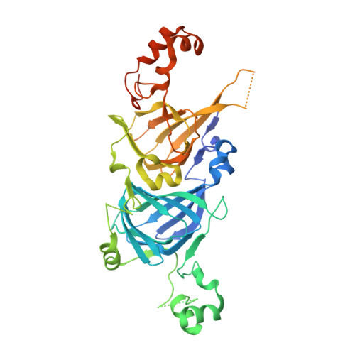
Crystal Structure of Adzuki Bean 7S Globulin-3


Experimental Data Snapshot

  • Method: X-RAY DIFFRACTION
  • Resolution: 2.25 Å
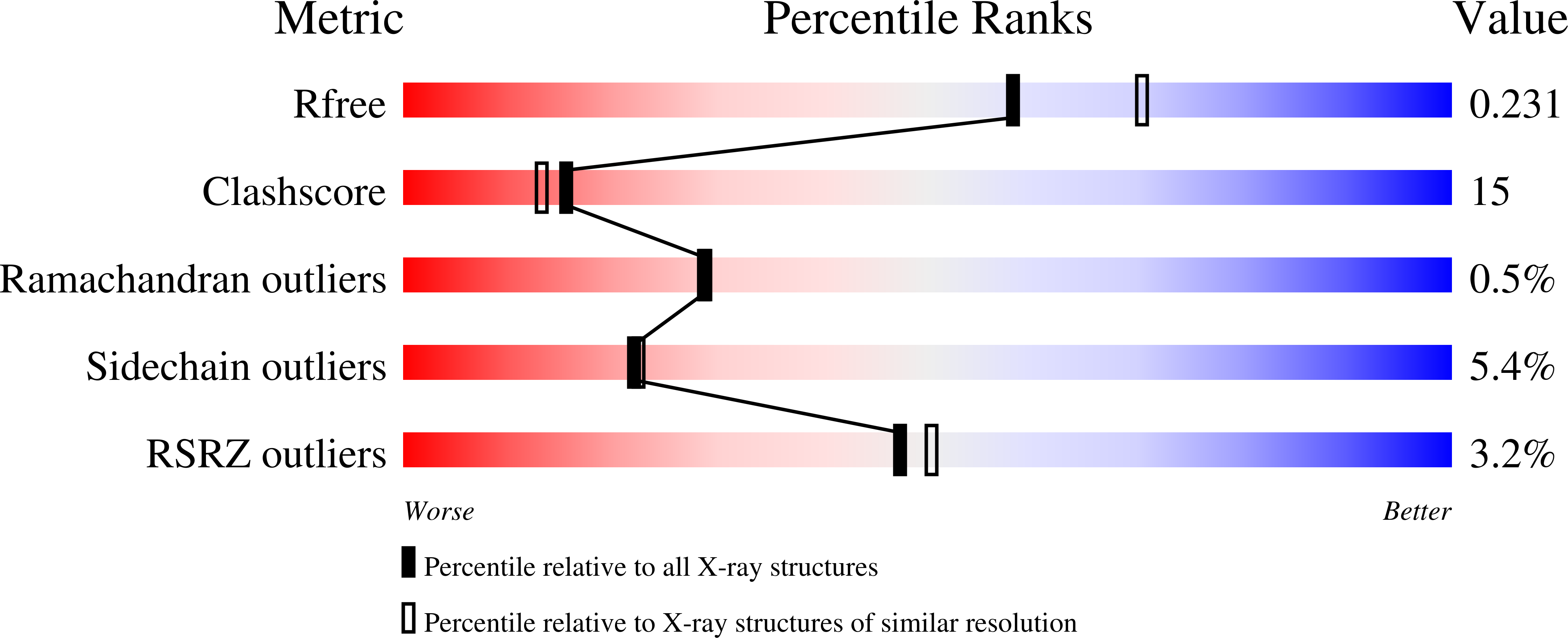
  • R-Value Free: 0.237 
  • R-Value Work: 0.194 
  • R-Value Observed: 0.194 

Starting Model: experimental
View more details

wwPDB Validation   3D Report Full Report


This is version 1.2 of the entry. See complete history


Literature

Characterization and crystallography of recombinant 7S globulins of Adzuki bean and structure-function relationships with 7S globulins of various crops.

Fukuda, T.Maruyama, N.Salleh, M.R.Mikami, B.Utsumi, S.

(2008) J Agric Food Chem 56: 4145-4153

  • DOI: https://doi.org/10.1021/jf072667b
  • Primary Citation of Related Structures:  
    2EA7, 2EAA

  • PubMed Abstract: 

    The recombinant proteins Adzuki 7S1, Adzuki 7S2, and Adzuki 7S3 were prepared through the Escherichia coli expression systems of three kinds of adzuki bean cDNAs. The recombinant proteins exhibited intrinsic thermal stabilities, surface hydrophobicities, and solubilities, although the homology of their amino acid sequences ranged from 95-98%. To understand why these individual proteins exhibited different properties, their three-dimensional structures were elucidated. The three proteins were successfully crystallized, and the three-dimensional structures of Adzuki 7S1 and Adzuki 7S3 were determined. The properties and structures of these two proteins were comprehensively compared with those of recombinant 7S globulins (soybean beta-conglycinins beta and alpha'c and mungbean 8Salpha) reported previously. It was likely that cavity sizes, hydrogen bonds, salt bridges, hydrophobic interactions, and lengths of loops determine the thermal stabilities of 7S globulins, and results indicated that cavity sizes strongly contribute to such stability. Surface hydrophobicity was also found to be determined not only by distributions of hydrophobic residues on the molecular surface. Furthermore, solubility at neutral and weak alkaline pH values at mu = 0.08 was found to be dominantly influenced by the electrostatic surface potentials.


  • Organizational Affiliation

    Laboratory of Food Quality Design and Development, Graduate School of Agriculture, Kyoto University, Kyoto, Japan.


Macromolecules
Find similar proteins by:  (by identity cutoff)  |  3D Structure
Entity ID: 1
MoleculeChains Sequence LengthOrganismDetailsImage
7S globulin-3
A, B, C
433Vigna angularisMutation(s): 0 
Gene Names: 7S-3
UniProt
Find proteins for A4PIA0 (Phaseolus angularis)
Explore A4PIA0 
Go to UniProtKB:  A4PIA0
Entity Groups  
Sequence Clusters30% Identity50% Identity70% Identity90% Identity95% Identity100% Identity
UniProt GroupA4PIA0
Sequence Annotations
Expand
  • Reference Sequence
Small Molecules
Ligands 3 Unique
IDChains Name / Formula / InChI Key2D Diagram3D Interactions
CIT
Query on CIT

Download Ideal Coordinates CCD File 
E [auth A],
H [auth B],
K [auth C]
CITRIC ACID
C6 H8 O7
KRKNYBCHXYNGOX-UHFFFAOYSA-N
ACY
Query on ACY

Download Ideal Coordinates CCD File 
F [auth A],
I [auth B],
L [auth C]
ACETIC ACID
C2 H4 O2
QTBSBXVTEAMEQO-UHFFFAOYSA-N
CA
Query on CA

Download Ideal Coordinates CCD File 
D [auth A],
G [auth B],
J [auth C]
CALCIUM ION
Ca
BHPQYMZQTOCNFJ-UHFFFAOYSA-N
Experimental Data & Validation

Experimental Data

  • Method: X-RAY DIFFRACTION
  • Resolution: 2.25 Å
  • R-Value Free: 0.237 
  • R-Value Work: 0.194 
  • R-Value Observed: 0.194 
  • Space Group: P 21 21 21
Unit Cell:
Length ( Å )Angle ( ˚ )
a = 66.873α = 90
b = 99.32β = 90
c = 216.868γ = 90
Software Package:
Software NamePurpose
CNSrefinement
HKL-2000data collection
HKL-2000data reduction
HKL-2000data scaling
CNSphasing

Structure Validation

View Full Validation Report


Entry History 

Deposition Data

Revision History  (Full details and data files)

  • Version 1.0: 2008-02-05
    Type: Initial release
  • Version 1.1: 2011-07-13
    Changes: Derived calculations, Version format compliance
  • Version 1.2: 2023-10-25
    Changes: Data collection, Database references, Derived calculations, Refinement description