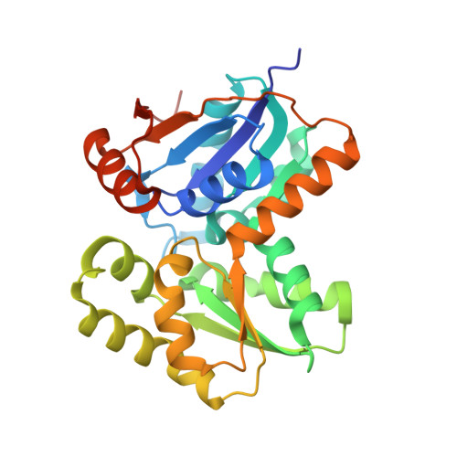
Structural and Functional Analysis of Two Glutamate Racemase Isozymes from Bacillus anthracis and Implications for Inhibitor Design
May, M., Mehboob, S., Mulhearn, D.C., Wang, Z., Yu, H., Thatcher, G.R.J., Santarsiero, B.D., Johnson, M.E., Mesecar, A.D.(2007) J Mol Biol 371: 1219-1237
- PubMed: 17610893
- DOI: https://doi.org/10.1016/j.jmb.2007.05.093
- Primary Citation of Related Structures:
2DWU, 2GZM - PubMed Abstract:
Glutamate racemase (RacE) is responsible for converting l-glutamate to d-glutamate, which is an essential component of peptidoglycan biosynthesis, and the primary constituent of the poly-gamma-d-glutamate capsule of the pathogen Bacillus anthracis. RacE enzymes are essential for bacterial growth and lack a human homolog, making them attractive targets for the design and development of antibacterial therapeutics. We have cloned, expressed and purified the two glutamate racemase isozymes, RacE1 and RacE2, from the B. anthracis genome. Through a series of steady-state kinetic studies, and based upon the ability of both RacE1 and RacE2 to catalyze the rapid formation of d-glutamate, we have determined that RacE1 and RacE2 are bona fide isozymes. The X-ray structures of B. anthracis RacE1 and RacE2, in complex with d-glutamate, were determined to resolutions of 1.75 A and 2.0 A. Both enzymes are dimers with monomers arranged in a "tail-to-tail" orientation, similar to the B. subtilis RacE structure, but differing substantially from the Aquifex pyrophilus RacE structure. The differences in quaternary structures produce differences in the active sites of racemases among the various species, which has important implications for structure-based, inhibitor design efforts within this class of enzymes. We found a Val to Ala variance at the entrance of the active site between RacE1 and RacE2, which results in the active site entrance being less sterically hindered for RacE1. Using a series of inhibitors, we show that this variance results in differences in the inhibitory activity against the two isozymes and suggest a strategy for structure-based inhibitor design to obtain broad-spectrum inhibitors for glutamate racemases.
Organizational Affiliation:
Center for Pharmaceutical Biotechnology, University of Illinois at Chicago, Chicago, IL 60607, USA.


