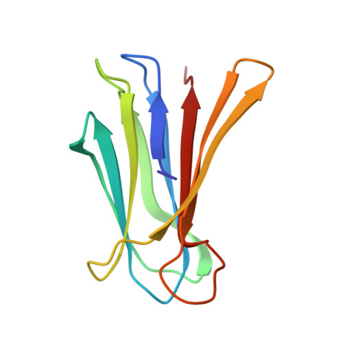
2BOJ

crystal Structure of pseudomonas aeruginosa lectin (PA-IIL) complexed with methyl-B-D-Arabinopyranoside


Experimental Data Snapshot

  • Method: X-RAY DIFFRACTION
  • Resolution: 1.80 Å
  • R-Value Free: 0.172 
  • R-Value Work: 0.136 
  • R-Value Observed: 0.137 

Starting Model: experimental
View more details

wwPDB Validation   3D Report Full Report


Ligand Structure Quality Assessment 


This is version 1.4 of the entry. See complete history


Literature

Binding of Different Monosaccharides by Lectin Pa-Iil from Pseudoman Aeruginosa: Thermodynamics Data Correlated with X-Ray Structures.

Sabin, C.D.Mitchell, E.P.Pokarna, M.Gautier, C.Utille, J.-P.Wimmerova, M.Imberty, A.

(2006) FEBS Lett 580: 982

  • DOI: https://doi.org/10.1016/j.febslet.2006.01.030
  • Primary Citation of Related Structures:  
    2BOJ, 2BP6

  • PubMed Abstract: 

    The lectin from Pseudomonas aeruginosa (PA-IIL) is involved in host recognition and biofilm formation. Lectin not only displays an unusually high affinity for fucose but also binds to L-fucose, L-galactose and D-arabinose that differ only by the group at position 5 of the sugar ring. Isothermal calorimetry experiments provided precise determination of affinity for the three methyl-glycosides and revealed a large enthalpy contribution. The crystal structures of the complexes of PA-IIL with L-galactose and Met-beta-D-arabinoside have been determined and compared with the PA-IIL/fucose complex described previously. A combination of the structures and thermodynamics provided clues for the role of the hydrophobic group in affinity.


  • Organizational Affiliation

    CERMAV-CNRS (affiliated with Université Joseph Fourier), 601 rue de la Chimie, Grenoble BP53, F-38041 Grenoble cedex 09, France.


Macromolecules
Find similar proteins by:  (by identity cutoff)  |  3D Structure
Entity ID: 1
MoleculeChains Sequence LengthOrganismDetailsImage
PSEUDOMONAS AERUGINOSA LECTIN II
A, B, C, D
114Pseudomonas aeruginosa PAO1Mutation(s): 0 
UniProt
Find proteins for Q9HYN5 (Pseudomonas aeruginosa (strain ATCC 15692 / DSM 22644 / CIP 104116 / JCM 14847 / LMG 12228 / 1C / PRS 101 / PAO1))
Explore Q9HYN5 
Go to UniProtKB:  Q9HYN5
Entity Groups  
Sequence Clusters30% Identity50% Identity70% Identity90% Identity95% Identity100% Identity
UniProt GroupQ9HYN5
Sequence Annotations
Expand
  • Reference Sequence
Small Molecules
Ligands 3 Unique
IDChains Name / Formula / InChI Key2D Diagram3D Interactions
ARW
Query on ARW

Download Ideal Coordinates CCD File 
E [auth A],
H [auth B],
K [auth C],
O [auth D]
methyl beta-D-arabinopyranoside
C6 H12 O5
ZBDGHWFPLXXWRD-ARQDHWQXSA-N
SO4
Query on SO4

Download Ideal Coordinates CCD File 
N [auth C]SULFATE ION
O4 S
QAOWNCQODCNURD-UHFFFAOYSA-L
CA
Query on CA

Download Ideal Coordinates CCD File 
F [auth A]
G [auth A]
I [auth B]
J [auth B]
L [auth C]
F [auth A],
G [auth A],
I [auth B],
J [auth B],
L [auth C],
M [auth C],
P [auth D],
Q [auth D]
CALCIUM ION
Ca
BHPQYMZQTOCNFJ-UHFFFAOYSA-N
Binding Affinity Annotations 
IDSourceBinding Affinity
ARW PDBBind:  2BOJ Kd: 1700 (nM) from 1 assay(s)
Experimental Data & Validation

Experimental Data

  • Method: X-RAY DIFFRACTION
  • Resolution: 1.80 Å
  • R-Value Free: 0.172 
  • R-Value Work: 0.136 
  • R-Value Observed: 0.137 
  • Space Group: P 1 21 1
Unit Cell:
Length ( Å )Angle ( ˚ )
a = 50.636α = 90
b = 80.168β = 109.92
c = 52.517γ = 90
Software Package:
Software NamePurpose
REFMACrefinement
DENZOdata reduction
SCALAdata scaling
AMoREphasing

Structure Validation

View Full Validation Report


Ligand Structure Quality Assessment 


Entry History 

Deposition Data

Revision History  (Full details and data files)

  • Version 1.0: 2006-02-22
    Type: Initial release
  • Version 1.1: 2011-05-08
    Changes: Version format compliance
  • Version 1.2: 2011-07-13
    Changes: Version format compliance
  • Version 1.3: 2020-07-29
    Type: Remediation
    Reason: Carbohydrate remediation
    Changes: Data collection, Derived calculations, Other, Structure summary
  • Version 1.4: 2023-12-13
    Changes: Data collection, Database references, Refinement description, Structure summary