2BJO

Crystal Structure of the Organic Hydroperoxide Resistance Protein OhrB of Bacillus subtilis


Experimental Data Snapshot

  • Method: X-RAY DIFFRACTION
  • Resolution: 2.10 Å
  • R-Value Free: 0.199 
  • R-Value Work: 0.158 
  • R-Value Observed: 0.161 

Starting Model: experimental
View more details

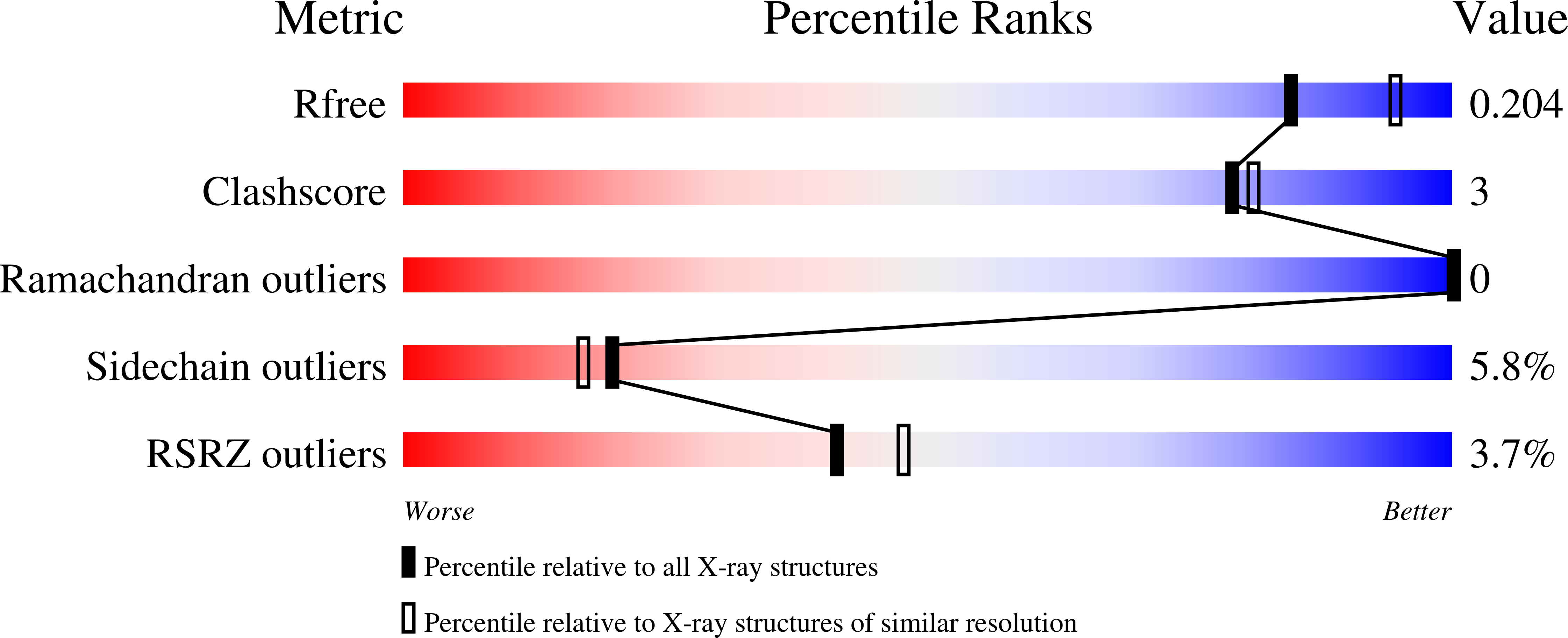
wwPDB Validation   3D Report Full Report


This is version 1.2 of the entry. See complete history


Literature

Structure of the Bacillus Subtilis Ohrb Hydroperoxide-Resistance Protein in a Fully Oxidized State.

Cooper, D.R.Surendranath, Y.Devedjiev, Y.Bielnicki, J.Derewenda, Z.S.

(2007) Acta Crystallogr D Biol Crystallogr 63: 1269

  • DOI: https://doi.org/10.1107/S0907444907050226
  • Primary Citation of Related Structures:  
    2BJO

  • PubMed Abstract: 

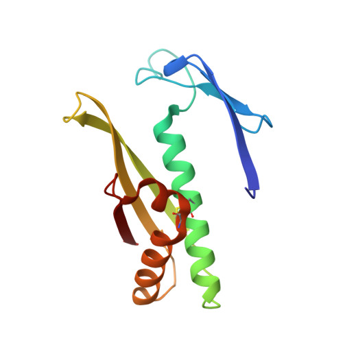
    The crystal structure of the fully oxidized form of the Bacillus subtilis organic hydroperoxide-resistance (OhrB) protein is reported at 2.1 A resolution. The electron density reveals an intact catalytic disulfide bond (Cys55-Cys119) in each of the two molecules, which are intertwined into a canonical obligate dimer. However, the stereochemistry of the disulfides is unorthodox and strained, suggesting that they are sensitive to reducing agents. A deep solvent-accessible gorge reaching Cys55 may represent the access route for the reductant.


  • Organizational Affiliation

    Department of Molecular Physiology and Biological Physics, University of Virginia and the PSI2 Integrated Center for Structure-Function Innovation Charlottesville, Virginia 22908, USA.


Macromolecules
Find similar proteins by:  (by identity cutoff)  |  3D Structure
Entity ID: 1
MoleculeChains Sequence LengthOrganismDetailsImage
ORGANIC HYDROPEROXIDE RESISTANCE PROTEIN OHRB
A, B
136Bacillus subtilisMutation(s): 3 
UniProt
Find proteins for P80242 (Bacillus subtilis (strain 168))
Explore P80242 
Go to UniProtKB:  P80242
Entity Groups  
Sequence Clusters30% Identity50% Identity70% Identity90% Identity95% Identity100% Identity
UniProt GroupP80242
Sequence Annotations
Expand
  • Reference Sequence
Experimental Data & Validation

Experimental Data

  • Method: X-RAY DIFFRACTION
  • Resolution: 2.10 Å
  • R-Value Free: 0.199 
  • R-Value Work: 0.158 
  • R-Value Observed: 0.161 
  • Space Group: P 1
Unit Cell:
Length ( Å )Angle ( ˚ )
a = 35.34α = 84.45
b = 41.434β = 91.51
c = 44.6γ = 73.89
Software Package:
Software NamePurpose
REFMACrefinement
HKL-2000data reduction
HKL-2000data scaling
AMoREphasing

Structure Validation

View Full Validation Report


Entry History 

Deposition Data

Revision History  (Full details and data files)

  • Version 1.0: 2006-03-09
    Type: Initial release
  • Version 1.1: 2011-07-13
    Changes: Advisory, Version format compliance
  • Version 1.2: 2023-12-13
    Changes: Data collection, Database references, Other, Refinement description