2B3K

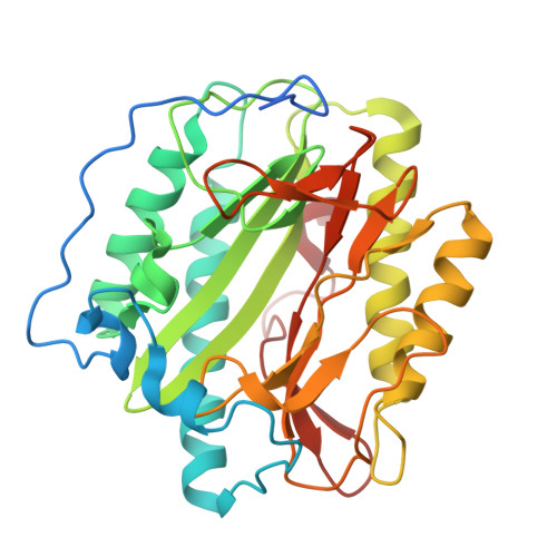
Crystal structure of Human Methionine Aminopeptidase Type I in the holo form


Experimental Data Snapshot

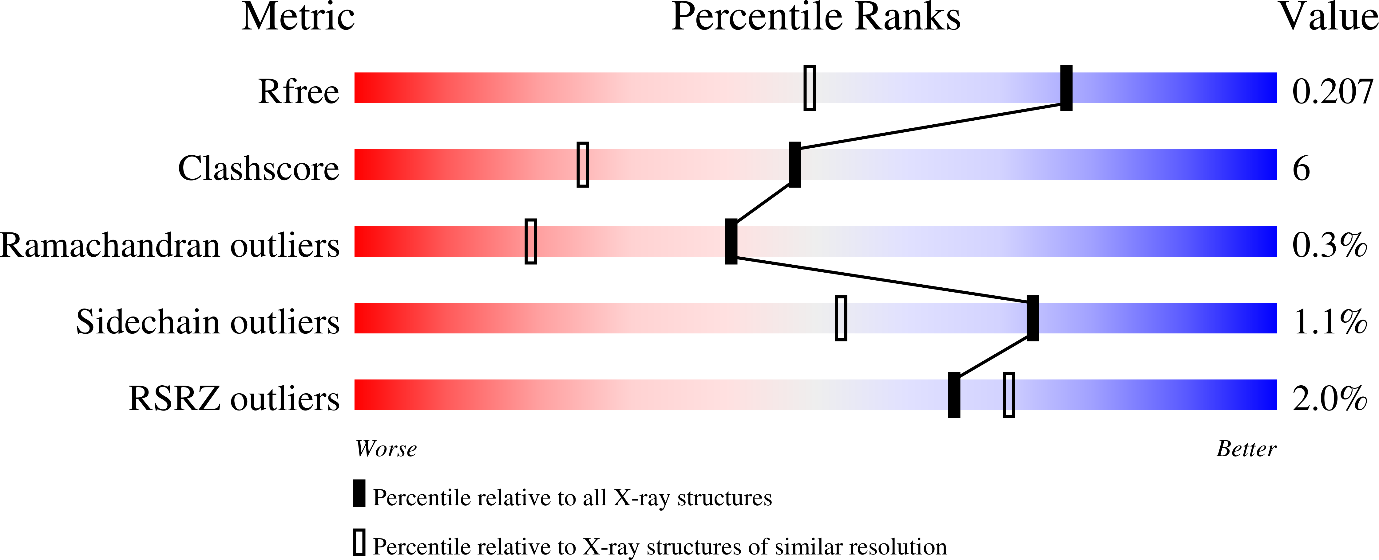
  • Method: X-RAY DIFFRACTION
  • Resolution: 1.55 Å
  • R-Value Free: 0.215 
  • R-Value Work: 0.190 
  • R-Value Observed: 0.190 

Starting Model: experimental
View more details

wwPDB Validation   3D Report Full Report


This is version 1.5 of the entry. See complete history

Re-refinement Note

A newer entry is available that reflects an alternative modeling of the original data: 8P2K


Literature

Structural Basis for the Functional Differences between Type I and Type II Human Methionine Aminopeptidases(,).

Addlagatta, A.Hu, X.Liu, J.O.Matthews, B.W.

(2005) Biochemistry 44: 14741-14749

  • DOI: https://doi.org/10.1021/bi051691k
  • Primary Citation of Related Structures:  
    2B3H, 2B3K, 2B3L

  • PubMed Abstract: 

    Determination of the crystal structure of human MetAP1 makes it possible, for the first time, to compare the structures of a Type I and a Type II methionine aminopeptidase (MetAP) from the same organism. Comparison of the Type I enzyme with the previously reported complex of ovalicin with Type II MetAP shows that the active site of the former is reduced in size and would incur steric clashes with the bound inhibitor. This explains why ovalicin and related anti-angiogenesis inhibitors target Type II human MetAP but not Type I. The differences in both size and shape of the active sites between MetAP1 and MetAP2 also help to explain their different substrate specificity. In the presence of excess Co(2+), a third cobalt ion binds in the active site region, explaining why metal ions in excess can be inhibitory. Also, the N-terminal region of the protein contains three distinct Pro-x-x-Pro motifs, supporting the prior suggestion that this region of the protein may participate in binding to the ribosome.


  • Organizational Affiliation

    Institute of Molecular Biology, Howard Hughes Medical Institute and Department of Physics, 1229 University of Oregon, Eugene, Oregon 97403-1229, USA.


Macromolecules
Find similar proteins by:  (by identity cutoff)  |  3D Structure
Entity ID: 1
MoleculeChains Sequence LengthOrganismDetailsImage
Methionine aminopeptidase 1329Homo sapiensMutation(s): 0 
Gene Names: METAP1KIAA0094
EC: 3.4.11.18
UniProt & NIH Common Fund Data Resources
Find proteins for P53582 (Homo sapiens)
Explore P53582 
Go to UniProtKB:  P53582
PHAROS:  P53582
GTEx:  ENSG00000164024 
Entity Groups  
Sequence Clusters30% Identity50% Identity70% Identity90% Identity95% Identity100% Identity
UniProt GroupP53582
Sequence Annotations
Expand
  • Reference Sequence
Experimental Data & Validation

Experimental Data

  • Method: X-RAY DIFFRACTION
  • Resolution: 1.55 Å
  • R-Value Free: 0.215 
  • R-Value Work: 0.190 
  • R-Value Observed: 0.190 
  • Space Group: P 1 21 1
Unit Cell:
Length ( Å )Angle ( ˚ )
a = 47.432α = 90
b = 77.522β = 90.6
c = 48.308γ = 90
Software Package:
Software NamePurpose
DENZOdata reduction
SCALEPACKdata scaling
EPMRphasing
CNSrefinement
PDB_EXTRACTdata extraction
DCREDUCEdata reduction

Structure Validation

View Full Validation Report


Entry History 

Deposition Data

Revision History  (Full details and data files)

  • Version 1.0: 2005-11-22
    Type: Initial release
  • Version 1.1: 2008-05-01
    Changes: Version format compliance
  • Version 1.2: 2011-07-13
    Changes: Non-polymer description, Version format compliance
  • Version 1.3: 2017-10-11
    Changes: Refinement description
  • Version 1.4: 2019-11-20
    Changes: Database references
  • Version 1.5: 2023-08-23
    Changes: Data collection, Database references, Derived calculations, Refinement description