Three-dimensional structure of neuropeptide k bound to dodecylphosphocholine micelles
Dike, A., Cowsik, S.M.(2006) Biochemistry 45: 2994-3004
- PubMed: 16503654
- DOI: https://doi.org/10.1021/bi052287o
- Primary Citation of Related Structures:
2B19 - PubMed Abstract:
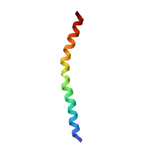
Neuropeptide K (NPK), an N-terminally extended form of neurokinin A (NKA), represents the most potent and longest lasting vasodepressor and cardiomodulatory tachykinin reported thus far. NPK has been shown to have high selectivity for the NK2 receptor. Because the micelle-associated structure may be relevant to the NPK-receptor interaction, the three-dimensional structure of the NPK in aqueous and micellar environments has been studied by two-dimensional proton nuclear magnetic resonance (2D (1)H NMR spectroscopy) and distance geometry calculations. Proton NMR assignments have been carried out with the aid of correlation spectroscopy (DQF-COSY and TOCSY) and nuclear Overhauser effect spectroscopy (NOESY and ROESY) experiments. The interproton distances and dihedral angle constraints obtained from the NMR data have been used in torsion angle dynamics algorithm for NMR applications (DYANA) to generate a family of structures, which have been refined using restrained energy minimization and dynamics. The results show that in an aqueous environment NPK lacks a definite secondary structure, although some turn-like elements are present in the N terminus. The structure is well-defined in the presence of dodecylphosphocholine micelles. The global fold of NPK bound to DPC micelles consists of two well-defined helices from residues 9 to 18 and residues 27 to 33 connected by a noncanonical beta turn. The N terminus of the peptide is characterized by a 3(10) helix or a series of dynamic beta turns. The conformational range of the peptide revealed by NMR and circular dichroism (CD) studies has been analyzed in terms of characteristic secondary features. The observed conformational features have been further compared to a NKA and neuropeptide gamma (NPgamma) potent endogenous agonist for the NK2 receptor.
Organizational Affiliation:
School of Life Sciences, Jawaharlal Nehru University, New Delhi 110067, India.