Structure and Dynamics of Micelle-Associated Human Immunodeficiency Virus gp41 Fusion Domain.
Jaroniec, C.P., Kaufman, J.D., Stahl, S.J., Viard, M., Blumenthal, R., Wingfield, P.T., Bax, A.(2005) Biochemistry 44: 16167-16180
- PubMed: 16331977
- DOI: https://doi.org/10.1021/bi051672a
- Primary Citation of Related Structures:
2ARI - PubMed Abstract:
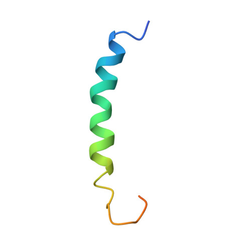
The N-terminal fusion domain of the HIV-1 gp41 envelope glycoprotein is responsible for initiating the fusion of viral and cellular membranes, leading to the subsequent infection of the host cell by HIV-1. We have investigated the backbone structure and dynamics of the 30 N-terminal residues of HIV-1 gp41 in membrane-mimicking environments using NMR spectroscopy and (15)N- and (15)N,(13)C,(2)H-labeled peptides. Similar (15)N-(1)H HSQC spectra were obtained in a variety of detergents, including SDS, DPC, mixed DPC/SDS, and LPPG micelles, indicating that the peptide structure is not strongly influenced by the type of detergent used. Detailed characterization was carried out in SDS micelles, where the long-term sample stability was found to be optimal. In addition to J-coupling and NOE restraints, a nearly complete set of backbone residual dipolar coupling restraints was recorded for the fusion domain-micelle complex aligned with respect to the magnetic field using a stretched polyacrylamide gel. Backbone amide (15)N spin relaxation and amide hydrogen exchange rates with the solvent were also measured. The ensemble of NMR structures reveals an uninterrupted alpha-helix for the least mobile residues (S(2) > 0.65), Ile-4 to Met-19, with transient helical character extending up to Ala-22. A 12-residue (Ile-4 to Ala-15) segment is fully shielded from solvent, with Gly-3 and Gly-16 found at micelle-solvent interfaces. Residues external to the micelle exhibit enhanced picosecond to nanosecond time scale dynamics relative to the residues buried in the micelle, and their mobility increases with the distance from the micelle.
Organizational Affiliation:
Laboratory of Chemical Physics, National Institute of Diabetes and Digestive and Kidney Diseases, National Institutes of Health, Bethesda, Maryland 20892-0520, USA. jaroniec@speck.niddk.nih.gov