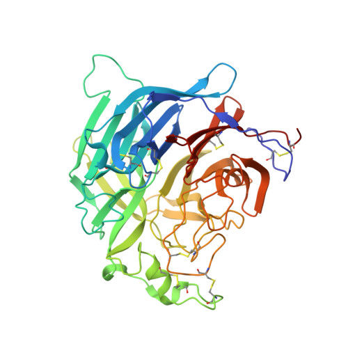
2X9M

Hendra virus attachment glycoprotein


Experimental Data Snapshot

  • Method: X-RAY DIFFRACTION
  • Resolution: 2.90 Å
  • R-Value Free: 0.273 
  • R-Value Work: 0.233 
  • R-Value Observed: 0.235 

Starting Model: experimental
View more details

wwPDB Validation   3D Report Full Report


Ligand Structure Quality Assessment 


This is version 1.5 of the entry. See complete history


Literature

Dimeric Architecture of the Hendra Virus Attachment Glycoprotein: Evidence for a Conserved Mode of Assembly.

Bowden, T.A.Crispin, M.Harvey, D.J.Jones, E.Y.Stuart, D.I.

(2010) J Virol 84: 6208

  • DOI: https://doi.org/10.1128/JVI.00317-10
  • Primary Citation of Related Structures:  
    2X9M

  • PubMed Abstract: 

    Hendra virus is a negative-sense single-stranded RNA virus within the Paramyxoviridae family which, together with Nipah virus, forms the Henipavirus genus. Infection with bat-borne Hendra virus leads to a disease with high mortality rates in humans. We determined the crystal structure of the unliganded six-bladed beta-propeller domain and compared it to the previously reported structure of Hendra virus attachment glycoprotein (HeV-G) in complex with its cellular receptor, ephrin-B2. As observed for the related unliganded Nipah virus structure, there is plasticity in the Glu579-Pro590 and Lys236-Ala245 ephrin-binding loops prior to receptor engagement. These data reveal that henipaviral attachment glycoproteins undergo common structural transitions upon receptor binding and further define the structural template for antihenipaviral drug design. Our analysis also provides experimental evidence for a dimeric arrangement of HeV-G that exhibits striking similarity to those observed in crystal structures of related paramyxovirus receptor-binding glycoproteins. The biological relevance of this dimer is further supported by the positional analysis of glycosylation sites from across the paramyxoviruses. In HeV-G, the sites lie away from the putative dimer interface and remain accessible to alpha-mannosidase processing on oligomerization. We therefore propose that the overall mode of dimer assembly is conserved for all paramyxoviruses; however, while the geometry of dimerization is rather closely similar for those viruses that bind flexible glycan receptors, significant (up to 60 degrees ) and different reconfigurations of the subunit packing (associated with a significant decrease in the size of the dimer interface) have accompanied the independent switching to high-affinity protein receptor binding in Hendra and measles viruses.


  • Organizational Affiliation

    Division of Structural Biology, Wellcome Trust Centre for Human Genetics, University of Oxford, Roosevelt Drive, Oxford OX3 7BN, United Kingdom.


Macromolecules
Find similar proteins by:  (by identity cutoff)  |  3D Structure
Entity ID: 1
MoleculeChains Sequence LengthOrganismDetailsImage
GLYCOPROTEIN G
A, B, C, D
420Henipavirus hendraenseMutation(s): 0 
UniProt
Find proteins for O89343 (Hendra virus (isolate Horse/Autralia/Hendra/1994))
Explore O89343 
Go to UniProtKB:  O89343
Entity Groups  
Sequence Clusters30% Identity50% Identity70% Identity90% Identity95% Identity100% Identity
UniProt GroupO89343
Glycosylation
Glycosylation Sites: 4
Sequence Annotations
Expand
  • Reference Sequence
Small Molecules
Ligands 1 Unique
IDChains Name / Formula / InChI Key2D Diagram3D Interactions
NAG
Query on NAG

Download Ideal Coordinates CCD File 
E [auth A]
F [auth A]
G [auth A]
H [auth A]
I [auth B]
E [auth A],
F [auth A],
G [auth A],
H [auth A],
I [auth B],
J [auth B],
K [auth B],
L [auth B],
M [auth C],
N [auth D]
2-acetamido-2-deoxy-beta-D-glucopyranose
C8 H15 N O6
OVRNDRQMDRJTHS-FMDGEEDCSA-N
Experimental Data & Validation

Experimental Data

  • Method: X-RAY DIFFRACTION
  • Resolution: 2.90 Å
  • R-Value Free: 0.273 
  • R-Value Work: 0.233 
  • R-Value Observed: 0.235 
  • Space Group: P 1
Unit Cell:
Length ( Å )Angle ( ˚ )
a = 65.883α = 103.39
b = 84.775β = 99.63
c = 91.548γ = 108.62
Software Package:
Software NamePurpose
REFMACrefinement
DENZOdata reduction
SCALEPACKdata scaling
PHASERphasing

Structure Validation

View Full Validation Report


Ligand Structure Quality Assessment 


Entry History 

Deposition Data

Revision History  (Full details and data files)

  • Version 1.0: 2010-04-21
    Type: Initial release
  • Version 1.1: 2011-05-08
    Changes: Version format compliance
  • Version 1.2: 2011-07-13
    Changes: Version format compliance
  • Version 1.3: 2020-07-29
    Type: Remediation
    Reason: Carbohydrate remediation
    Changes: Data collection, Derived calculations, Other, Structure summary
  • Version 1.4: 2023-12-20
    Changes: Data collection, Database references, Refinement description, Structure summary
  • Version 1.5: 2024-11-06
    Changes: Structure summary