The Glutamate Receptor Glur5 Agonist (S)-2-Amino-3-(3-Hydroxy-7,8-Dihydro-6H-Cyclohepta[D]Isoxazol-4-Yl)Propionic Acid and the 8-Methyl Analogue: Synthesis, Molecular Pharmacology, and Biostructural Characterization
Clausen, R.P., Naur, P., Kristensen, A.S., Greenwood, J.R., Strange, M., Brauner-Osborne, H., Jensen, A.A., Nielsen, A.S., Geneser, U., Ringgaard, L.M., Nielsen, B., Pickering, D.S., Brehm, L., Gajhede, M., Krogsgaard-Larsen, P., Kastrup, J.S.(2009) J Med Chem 52: 4911
- PubMed: 19588945
- DOI: https://doi.org/10.1021/jm900565c
- Primary Citation of Related Structures:
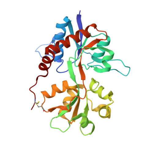
2WKY - PubMed Abstract:
The design, synthesis, and pharmacological characterization of a highly potent and selective glutamate GluR5 agonist is reported. (S)-2-Amino-3-((RS)-3-hydroxy-8-methyl-7,8-dihydro-6H-cyclohepta[d]isoxazol-4-yl)propionic acid (5) is the 8-methyl analogue of (S)-2-amino-3-(3-hydroxy-7,8-dihydro-6H-cyclohepta[d]isoxazol-4-yl)propionic acid ((S)-4-AHCP, 4). Compound 5 displays an improved selectivity profile compared to 4. A versatile stereoselective synthetic route for this class of compounds is presented along with the characterization of the binding affinity of 5 to ionotropic glutamate receptors (iGluRs). Functional characterization of 5 at cloned iGluRs using a calcium imaging assay and voltage-clamp recordings show a different activation of GluR5 compared to (S)-glutamic acid (Glu), kainic acid (KA, 1), and (S)-2-amino-3-(3-hydroxy-5-tert-butyl-4-isoxazolyl)propionic acid ((S)-ATPA, 3) as previously demonstrated for 4. An X-ray crystallographic analysis of 4 and computational analyses of 4 and 5 bound to the GluR5 agonist binding domain (ABD) are presented, including a watermap analysis, which suggests that water molecules in the agonist binding site are important selectivity determinants.
Organizational Affiliation:
Department of Medicinal Chemistry, Faculty of Pharmaceutical Sciences, University of Copenhagen, 2 Universitetsparken, DK-2100 Copenhagen, Denmark. rac@farma.ku.dk