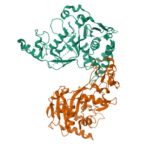
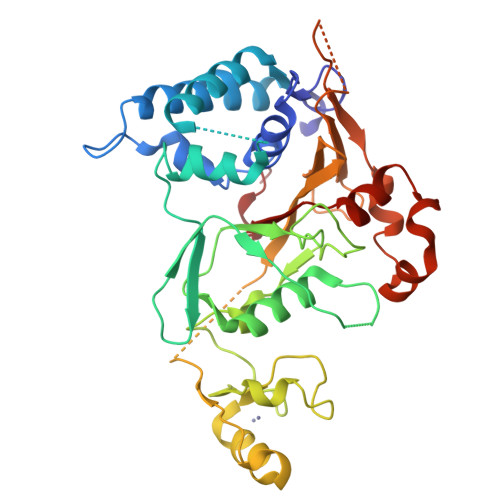
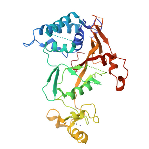
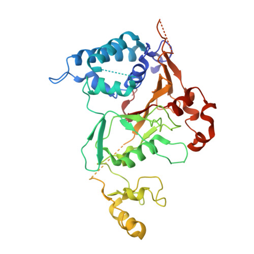
2VHF

Structure of the CYLD USP domain


Experimental Data Snapshot

  • Method: X-RAY DIFFRACTION
  • Resolution: 2.80 Å
  • R-Value Free: 0.281 
  • R-Value Work: 0.233 
  • R-Value Observed: 0.235 

wwPDB Validation   3D Report Full Report


This is version 1.3 of the entry. See complete history


Literature

The Structure of the Cyld Usp Domain Explains its Specificity for Lys63-Linked Polyubiquitin and Reveals a B-Box Module

Komander, D.Lord, C.J.Scheel, H.Swift, S.Hofmann, K.Ashworth, A.Barford, D.

(2008) Mol Cell Biol 29: 451

  • DOI: https://doi.org/10.1016/j.molcel.2007.12.018
  • Primary Citation of Related Structures:  
    2VHF

  • PubMed Abstract: 

    The tumor suppressor CYLD antagonizes NF-kappaB and JNK signaling by disassembly of Lys63-linked ubiquitin chains synthesized in response to cytokine stimulation. Here we describe the crystal structure of the CYLD USP domain, revealing a distinctive architecture that provides molecular insights into its specificity toward Lys63-linked polyubiquitin. We identify regions of the USP domain responsible for this specificity and demonstrate endodeubiquitinase activity toward such chains. Pathogenic truncations of the CYLD C terminus, associated with the hypertrophic skin tumor cylindromatosis, disrupt the USP domain, accounting for loss of CYLD catalytic activity. A small zinc-binding B box domain, similar in structure to other crossbrace Zn-binding folds--including the RING domain found in E3 ubiquitin ligases--is inserted within the globular core of the USP domain. Biochemical and functional characterization of the B box suggests a role as a protein-interaction module that contributes to determining the subcellular localization of CYLD.


  • Organizational Affiliation

    Section of Structural Biology, Institute of Cancer Research, Chester Beatty Laboratories, 237 Fulham Road, London SW3 6JB, UK.


Macromolecules
Find similar proteins by:  (by identity cutoff)  |  3D Structure
Entity ID: 1
MoleculeChains Sequence LengthOrganismDetailsImage
UBIQUITIN CARBOXYL-TERMINAL HYDROLASE CYLD
A, B
374Homo sapiensMutation(s): 0 
EC: 3.1.2.15 (PDB Primary Data), 3.4.19.12 (UniProt)
UniProt & NIH Common Fund Data Resources
Find proteins for Q9NQC7 (Homo sapiens)
Explore Q9NQC7 
Go to UniProtKB:  Q9NQC7
PHAROS:  Q9NQC7
GTEx:  ENSG00000083799 
Entity Groups  
Sequence Clusters30% Identity50% Identity70% Identity90% Identity95% Identity100% Identity
UniProt GroupQ9NQC7
Sequence Annotations
Expand
  • Reference Sequence
Experimental Data & Validation

Experimental Data

  • Method: X-RAY DIFFRACTION
  • Resolution: 2.80 Å
  • R-Value Free: 0.281 
  • R-Value Work: 0.233 
  • R-Value Observed: 0.235 
  • Space Group: P 21 21 21
Unit Cell:
Length ( Å )Angle ( ˚ )
a = 60.494α = 90
b = 89.083β = 90
c = 171.806γ = 90
Software Package:
Software NamePurpose
REFMACrefinement
MOSFLMdata reduction
SCALAdata scaling
SHELXCDEphasing
HKL2Mapphasing
SHARPphasing

Structure Validation

View Full Validation Report


Entry History 

Deposition Data

Revision History  (Full details and data files)

  • Version 1.0: 2008-03-11
    Type: Initial release
  • Version 1.1: 2011-05-08
    Changes: Version format compliance
  • Version 1.2: 2011-07-13
    Changes: Version format compliance
  • Version 1.3: 2024-05-08
    Changes: Data collection, Database references, Derived calculations, Other, Refinement description