2V64

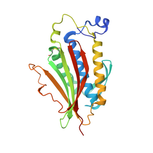
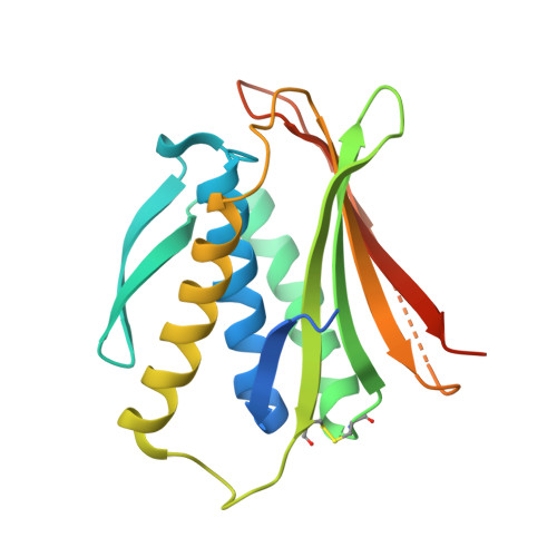
Crystallographic structure of the conformational dimer of the Spindle Assembly Checkpoint protein Mad2.


Experimental Data Snapshot

  • Method: X-RAY DIFFRACTION
  • Resolution: 2.90 Å
  • R-Value Free: 0.273 
  • R-Value Work: 0.233 
  • R-Value Observed: 0.233 

Starting Model: experimental
View more details

wwPDB Validation   3D Report Full Report


This is version 1.3 of the entry. See complete history


Literature

The MAD2 Conformational Dimer: Structure and Implications for the Spindle Assembly Checkpoint

Mapelli, M.Massimiliano, L.Santaguida, S.Musacchio, A.

(2007) Cell 131: 730

  • DOI: https://doi.org/10.1016/j.cell.2007.08.049
  • Primary Citation of Related Structures:  
    2V64

  • PubMed Abstract: 

    The 25 kDa Mad2 protein is a key player in the spindle assembly checkpoint, a safeguard against chromosome segregation errors in mitosis. Mad2 combines three unusual properties. First, Mad2 adopts two conformations with distinct topologies, open (O) and closed (C) Mad2. Second, C-Mad2 forms topological links with its two best-characterized protein ligands, Mad1 and Cdc20. Third, O-Mad2 and C-Mad2 engage in a "conformational" dimer that is essential for spindle checkpoint function in different organisms. The crystal structure of the O-Mad2-C-Mad2 conformational dimer, reported here, reveals an asymmetric interface that explains the selective dimerization of the O-Mad2 and C-Mad2 conformers. The structure also identifies several buried hydrophobic residues whose rearrangement correlates with the Mad2 topological change. The structure of the O-Mad2-C-Mad2 conformational dimer is consistent with a catalytic model in which a C-Mad2 template facilitates the binding of O-Mad2 to Cdc20, the target of Mad2 in the spindle checkpoint.


  • Organizational Affiliation

    Department of Experimental Oncology, European Institute of Oncology, Via Adamello 16, I-20139, Milan, Italy. marina.mapelli@ifom-ieo-campus.it


Macromolecules
Find similar proteins by:  (by identity cutoff)  |  3D Structure
Entity ID: 1
MoleculeChains Sequence LengthOrganismDetailsImage
MITOTIC SPINDLE ASSEMBLY CHECKPOINT PROTEIN MAD2A
A, C, F
213Homo sapiensMutation(s): 0 
UniProt & NIH Common Fund Data Resources
Find proteins for Q13257 (Homo sapiens)
Explore Q13257 
Go to UniProtKB:  Q13257
PHAROS:  Q13257
GTEx:  ENSG00000164109 
Entity Groups  
Sequence Clusters30% Identity50% Identity70% Identity90% Identity95% Identity100% Identity
UniProt GroupQ13257
Sequence Annotations
Expand
  • Reference Sequence

Find similar proteins by:  Sequence   |   3D Structure  

Entity ID: 2
MoleculeChains Sequence LengthOrganismDetailsImage
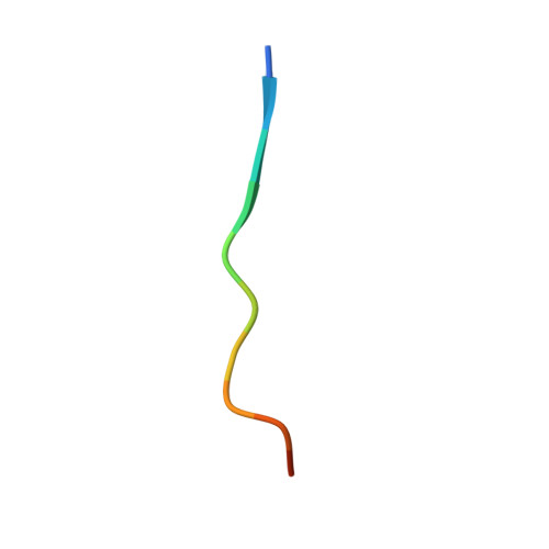
MBP1
B, G, I
12synthetic constructMutation(s): 0 
Entity Groups  
Sequence Clusters30% Identity50% Identity70% Identity90% Identity95% Identity100% Identity
Sequence Annotations
Expand
  • Reference Sequence
Find similar proteins by:  (by identity cutoff)  |  3D Structure
Entity ID: 3
MoleculeChains Sequence LengthOrganismDetailsImage
MITOTIC SPINDLE ASSEMBLY CHECKPOINT PROTEIN MAD2A
D, E, H
207Homo sapiensMutation(s): 0 
UniProt & NIH Common Fund Data Resources
Find proteins for Q13257 (Homo sapiens)
Explore Q13257 
Go to UniProtKB:  Q13257
PHAROS:  Q13257
GTEx:  ENSG00000164109 
Entity Groups  
Sequence Clusters30% Identity50% Identity70% Identity90% Identity95% Identity100% Identity
UniProt GroupQ13257
Sequence Annotations
Expand
  • Reference Sequence
Experimental Data & Validation

Experimental Data

  • Method: X-RAY DIFFRACTION
  • Resolution: 2.90 Å
  • R-Value Free: 0.273 
  • R-Value Work: 0.233 
  • R-Value Observed: 0.233 
  • Space Group: P 21 21 21
Unit Cell:
Length ( Å )Angle ( ˚ )
a = 112.58α = 90
b = 111.88β = 90
c = 131.76γ = 90
Software Package:
Software NamePurpose
CNSrefinement
HKL-2000data reduction
HKL-2000data scaling
PHASERphasing

Structure Validation

View Full Validation Report


Entry History 

Deposition Data

Revision History  (Full details and data files)

  • Version 1.0: 2007-11-27
    Type: Initial release
  • Version 1.1: 2016-12-21
    Changes: Derived calculations, Non-polymer description, Other, Refinement description, Source and taxonomy, Version format compliance
  • Version 1.2: 2023-12-13
    Changes: Data collection, Database references, Other, Refinement description
  • Version 1.3: 2024-10-23
    Changes: Structure summary