2OZ4

Structural Plasticity in IgSF Domain 4 of ICAM-1 Mediates Cell Surface Dimerization


Experimental Data Snapshot

  • Method: X-RAY DIFFRACTION
  • Resolution: 2.70 Å
  • R-Value Free: 0.253 
  • R-Value Work: 0.204 
  • R-Value Observed: 0.209 

Starting Models: experimental
View more details

wwPDB Validation   3D Report Full Report


Ligand Structure Quality Assessment 


This is version 2.1 of the entry. See complete history


Literature

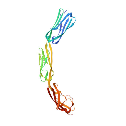
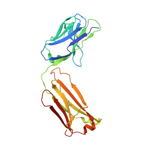
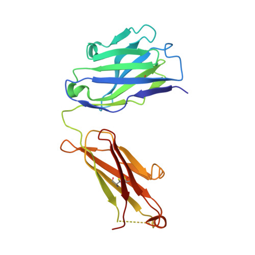
Structural plasticity in Ig superfamily domain 4 of ICAM-1 mediates cell surface dimerization.

Chen, X.Kim, T.D.Carman, C.V.Mi, L.Z.Song, G.Springer, T.A.

(2007) Proc Natl Acad Sci U S A 104: 15358-15363

  • DOI: https://doi.org/10.1073/pnas.0707406104
  • Primary Citation of Related Structures:  
    2OZ4

  • PubMed Abstract: 

    The Ig superfamily (IgSF) intercellular adhesion molecule-1 (ICAM-1) equilibrates between monomeric and dimeric forms on the cell surface, and dimerization enhances cell adhesion. A crystal structure of ICAM-1 IgSF domains (D) 3-5 revealed a unique dimerization interface in which D4s of two protomers fuse through edge beta-strands to form a single super beta-sandwich domain. Here, we describe a crystal structure at 2.7-A resolution of monomeric ICAM-1 D3-D5, stabilized by the monomer-specific Fab CA7. CA7 binds to D5 in a region that is buried in the dimeric interface and is distal from the dimerization site in D4. In monomeric ICAM-1 D3-D5, a 16-residue loop in D4 that is disordered in the dimeric structure could clearly be traced as a BC loop, a short C strand, and a CE meander with a cis-Pro followed by a solvent-exposed, flexible four-residue region. Deletions of 6 or 10 residues showed that the C-strand is essential for monomer stability, whereas a distinct six-residue deletion showed little contribution of the CE meander. Mutation of two inward-pointing Leu residues in edge beta-strand E to Lys increased monomer stability, confirming the hypothesis that inward-pointing charged side chains on edge beta-strands are an important design feature to prevent beta-supersheet formation. Overall, the studies reveal that monomer-dimer transition is associated with a surprisingly large, physiologically relevant, IgSF domain rearrangement.


  • Organizational Affiliation

    Immune Disease Institute, Department of Pathology, Harvard Medical School, Boston, MA 02115, USA.


Macromolecules
Find similar proteins by:  (by identity cutoff)  |  3D Structure
Entity ID: 1
MoleculeChains Sequence LengthOrganismDetailsImage
Intercellular adhesion molecule 1265Homo sapiensMutation(s): 0 
Gene Names: ICAM1
UniProt & NIH Common Fund Data Resources
Find proteins for P05362 (Homo sapiens)
Explore P05362 
Go to UniProtKB:  P05362
GTEx:  ENSG00000090339 
Entity Groups  
Sequence Clusters30% Identity50% Identity70% Identity90% Identity95% Identity100% Identity
UniProt GroupP05362
Glycosylation
Glycosylation Sites: 3Go to GlyGen: P05362-1
Sequence Annotations
Expand
  • Reference Sequence
Find similar proteins by:  (by identity cutoff)  |  3D Structure
Entity ID: 2
MoleculeChains Sequence LengthOrganismDetailsImage
FAB FRAGMENT LIGHT CHAINB [auth L]214Mus musculusMutation(s): 0 
Entity Groups  
Sequence Clusters30% Identity50% Identity70% Identity90% Identity95% Identity100% Identity
Sequence Annotations
Expand
  • Reference Sequence
Find similar proteins by:  (by identity cutoff)  |  3D Structure
Entity ID: 3
MoleculeChains Sequence LengthOrganismDetailsImage
FAB FRAGMENT, HEAVY CHAINC [auth H]214Mus musculusMutation(s): 0 
UniProt
Find proteins for P01757 (Mus musculus)
Explore P01757 
Go to UniProtKB:  P01757
Entity Groups  
Sequence Clusters30% Identity50% Identity70% Identity90% Identity95% Identity100% Identity
UniProt GroupP01757
Sequence Annotations
Expand
  • Reference Sequence
Oligosaccharides

Help

Entity ID: 4
MoleculeChains Length2D Diagram Glycosylation3D Interactions
alpha-L-fucopyranose-(1-6)-2-acetamido-2-deoxy-beta-D-glucopyranoseD [auth B]2N-Glycosylation
Glycosylation Resources
GlyTouCan:  G86851RC
GlyCosmos:  G86851RC
GlyGen:  G86851RC
Small Molecules
Ligands 4 Unique
IDChains Name / Formula / InChI Key2D Diagram3D Interactions
NAG
Query on NAG

Download Ideal Coordinates CCD File 
E [auth A],
F [auth A]
2-acetamido-2-deoxy-beta-D-glucopyranose
C8 H15 N O6
OVRNDRQMDRJTHS-FMDGEEDCSA-N
TRS
Query on TRS

Download Ideal Coordinates CCD File 
K [auth L],
L
2-AMINO-2-HYDROXYMETHYL-PROPANE-1,3-DIOL
C4 H12 N O3
LENZDBCJOHFCAS-UHFFFAOYSA-O
SO4
Query on SO4

Download Ideal Coordinates CCD File 
G [auth A],
H [auth L]
SULFATE ION
O4 S
QAOWNCQODCNURD-UHFFFAOYSA-L
ZN
Query on ZN

Download Ideal Coordinates CCD File 
I [auth L],
J [auth L]
ZINC ION
Zn
PTFCDOFLOPIGGS-UHFFFAOYSA-N
Experimental Data & Validation

Experimental Data

  • Method: X-RAY DIFFRACTION
  • Resolution: 2.70 Å
  • R-Value Free: 0.253 
  • R-Value Work: 0.204 
  • R-Value Observed: 0.209 
  • Space Group: C 1 2 1
Unit Cell:
Length ( Å )Angle ( ˚ )
a = 185.396α = 90
b = 69.342β = 112.76
c = 88.173γ = 90
Software Package:
Software NamePurpose
REFMACrefinement
HKL-2000data collection
HKL-2000data reduction
HKL-2000data scaling
MOLREPphasing

Structure Validation

View Full Validation Report


Ligand Structure Quality Assessment 


Entry History 

Deposition Data

Revision History  (Full details and data files)

  • Version 1.0: 2007-10-16
    Type: Initial release
  • Version 1.1: 2011-07-13
    Changes: Advisory, Version format compliance
  • Version 2.0: 2020-07-29
    Type: Remediation
    Reason: Carbohydrate remediation
    Changes: Atomic model, Data collection, Derived calculations, Structure summary
  • Version 2.1: 2023-08-30
    Changes: Data collection, Database references, Refinement description, Structure summary