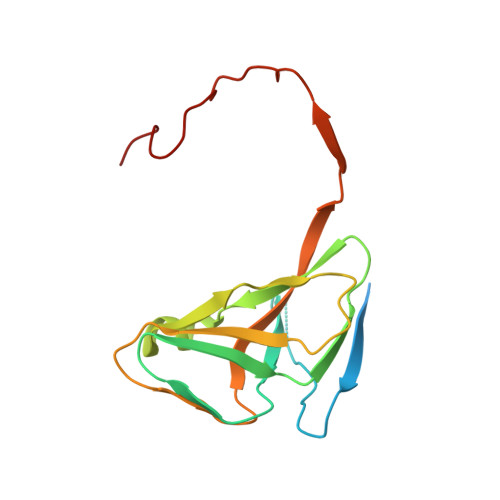
2HQU

Human dUTPase in complex with alpha,beta-iminodUTP and magnesium ion


Experimental Data Snapshot

  • Method: X-RAY DIFFRACTION
  • Resolution: 2.20 Å
  • R-Value Free: 0.251 
  • R-Value Work: 0.200 
  • R-Value Observed: 0.202 

Starting Model: experimental
View more details

wwPDB Validation   3D Report Full Report


Ligand Structure Quality Assessment 


This is version 1.4 of the entry. See complete history


Literature

Active site closure facilitates juxtaposition of reactant atoms for initiation of catalysis by human dUTPase.

Varga, B.Barabas, O.Kovari, J.Toth, J.Hunyadi-Gulyas, E.Klement, E.Medzihradszky, K.F.Tolgyesi, F.Fidy, J.Vertessy, B.G.

(2007) FEBS Lett 581: 4783-4788

  • DOI: https://doi.org/10.1016/j.febslet.2007.09.005
  • Primary Citation of Related Structures:  
    2HQU

  • PubMed Abstract: 

    Human dUTPase, essential for DNA integrity, is an important survival factor for cancer cells. We determined the crystal structure of the enzyme:alpha,beta-imino-dUTP:Mg complex and performed equilibrium binding experiments in solution. Ordering of the C-terminus upon the active site induces close juxtaposition of the incoming nucleophile attacker water oxygen and the alpha-phosphorus of the substrate, decreasing their distance below the van der Waals limit. Complex interactions of the C-terminus with both substrate and product were observed via a specifically designed tryptophan sensor, suitable for further detailed kinetic and ligand binding studies. Results explain the key functional role of the C-terminus.


  • Organizational Affiliation

    Institute of Enzymology, Biological Research Center, Hungarian Academy of Sciences, Budapest, Hungary.


Macromolecules
Find similar proteins by:  (by identity cutoff)  |  3D Structure
Entity ID: 1
MoleculeChains Sequence LengthOrganismDetailsImage
Deoxyuridine 5'-triphosphate nucleotidohydrolase
A, B, C
164Homo sapiensMutation(s): 0 
Gene Names: DUTN
EC: 3.6.1.23
UniProt & NIH Common Fund Data Resources
Find proteins for P33316 (Homo sapiens)
Explore P33316 
Go to UniProtKB:  P33316
PHAROS:  P33316
GTEx:  ENSG00000128951 
Entity Groups  
Sequence Clusters30% Identity50% Identity70% Identity90% Identity95% Identity100% Identity
UniProt GroupP33316
Sequence Annotations
Expand
  • Reference Sequence
Small Molecules
Ligands 3 Unique
IDChains Name / Formula / InChI Key2D Diagram3D Interactions
DUP
Query on DUP

Download Ideal Coordinates CCD File 
F [auth A],
H [auth B],
K [auth C]
2'-DEOXYURIDINE 5'-ALPHA,BETA-IMIDO-TRIPHOSPHATE
C9 H16 N3 O13 P3
XZLLMTSKYYYJLH-SHYZEUOFSA-N
CL
Query on CL

Download Ideal Coordinates CCD File 
J [auth C]CHLORIDE ION
Cl
VEXZGXHMUGYJMC-UHFFFAOYSA-M
MG
Query on MG

Download Ideal Coordinates CCD File 
D [auth A],
E [auth A],
G [auth B],
I [auth C]
MAGNESIUM ION
Mg
JLVVSXFLKOJNIY-UHFFFAOYSA-N
Binding Affinity Annotations 
IDSourceBinding Affinity
DUP PDBBind:  2HQU Kd: 7200 (nM) from 1 assay(s)
Experimental Data & Validation

Experimental Data

  • Method: X-RAY DIFFRACTION
  • Resolution: 2.20 Å
  • R-Value Free: 0.251 
  • R-Value Work: 0.200 
  • R-Value Observed: 0.202 
  • Space Group: P 21 21 21
Unit Cell:
Length ( Å )Angle ( ˚ )
a = 65.409α = 90
b = 71.773β = 90
c = 76.475γ = 90
Software Package:
Software NamePurpose
REFMACrefinement
XDSdata reduction
XSCALEdata scaling
MOLREPphasing

Structure Validation

View Full Validation Report


Ligand Structure Quality Assessment 


Entry History 

Deposition Data

Revision History  (Full details and data files)

  • Version 1.0: 2007-07-24
    Type: Initial release
  • Version 1.1: 2008-05-01
    Changes: Version format compliance
  • Version 1.2: 2011-07-13
    Changes: Advisory, Refinement description, Version format compliance
  • Version 1.3: 2016-10-19
    Changes: Refinement description
  • Version 1.4: 2023-08-30
    Changes: Data collection, Database references, Derived calculations, Refinement description