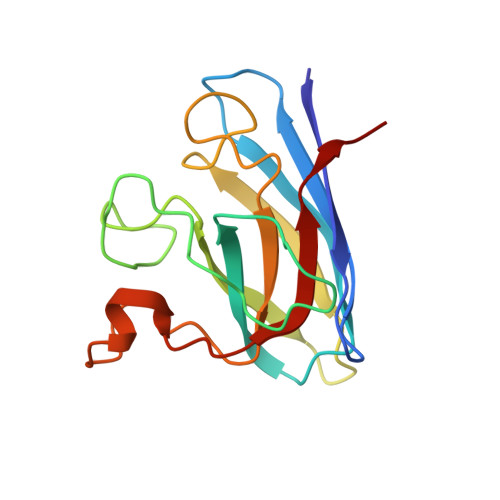
2GBU

C6A/C111A/C57A/C146A apo CuZn Superoxide dismutase


Experimental Data Snapshot

  • Method: X-RAY DIFFRACTION
  • Resolution: 2.00 Å
  • R-Value Free: 0.257 
  • R-Value Work: 0.217 
  • R-Value Observed: 0.219 

Starting Model: experimental
View more details

wwPDB Validation   3D Report Full Report


This is version 1.5 of the entry. See complete history


Literature

The Coupling between Disulphide Status, Metallation and Dimer Interface Strength in Cu/Zn Superoxide Dismutase

Hornberg, A.Logan, D.T.Marklund, S.L.Oliveberg, M.

(2007) J Mol Biol 365: 333-342

  • DOI: https://doi.org/10.1016/j.jmb.2006.09.048
  • Primary Citation of Related Structures:  
    2GBT, 2GBU, 2GBV

  • PubMed Abstract: 

    The gain of neurotoxic function in amyotrophic lateral sclerosis (ALS) has been linked to misfolding of the homodimeric enzyme Cu/Zn superoxide dismutase (SOD). Here, we present the crystal structure of fully cysteine-depleted human SOD (SOD(CallA)), representing a reduced, marginally stable intermediate on the folding pathway in vivo that has also been implicated as neurotoxic precursor state. A hallmark of this species is that it fails to dimerize and becomes trapped as a monomer in the absence of the active-site metals. The crystallographic data show that removal of the C57-C146 disulphide bond sets free the interface loop IV in the apo protein, whereas the same loop remains unaffected in the holo protein. Thus, the low dimerisation propensity of disulphide-reduced apoSOD seems to be of entropic origin due to increased loop flexibility in the monomeric state: in the disulphide-reduced holo protein this gain in configurational entropy upon splitting of the dimer interface is reduced by the metal coordination.


  • Organizational Affiliation

    Department of Biochemistry, Umeå University, SE-901 87 Umeå, Sweden.


Macromolecules
Find similar proteins by:  (by identity cutoff)  |  3D Structure
Entity ID: 1
MoleculeChains Sequence LengthOrganismDetailsImage
Superoxide dismutase [Cu-Zn]
A, B, C, D
153Homo sapiensMutation(s): 4 
Gene Names: SOD1
EC: 1.15.1.1
UniProt & NIH Common Fund Data Resources
Find proteins for P00441 (Homo sapiens)
Explore P00441 
Go to UniProtKB:  P00441
PHAROS:  P00441
GTEx:  ENSG00000142168 
Entity Groups  
Sequence Clusters30% Identity50% Identity70% Identity90% Identity95% Identity100% Identity
UniProt GroupP00441
Sequence Annotations
Expand
  • Reference Sequence
Experimental Data & Validation

Experimental Data

  • Method: X-RAY DIFFRACTION
  • Resolution: 2.00 Å
  • R-Value Free: 0.257 
  • R-Value Work: 0.217 
  • R-Value Observed: 0.219 
  • Space Group: C 1 2 1
Unit Cell:
Length ( Å )Angle ( ˚ )
a = 156.614α = 90
b = 34.427β = 112.17
c = 114.461γ = 90
Software Package:
Software NamePurpose
SCALAdata scaling
REFMACrefinement
PDB_EXTRACTdata extraction
MOSFLMdata reduction
CCP4data scaling
CNSphasing

Structure Validation

View Full Validation Report


Entry History 

Deposition Data

Revision History  (Full details and data files)

  • Version 1.0: 2007-01-02
    Type: Initial release
  • Version 1.1: 2008-05-01
    Changes: Version format compliance
  • Version 1.2: 2011-07-13
    Changes: Advisory, Version format compliance
  • Version 1.3: 2018-05-23
    Changes: Data collection
  • Version 1.4: 2021-11-10
    Changes: Database references
  • Version 1.5: 2023-10-25
    Changes: Data collection, Refinement description