2EB9

Crystal Structure of Cu(II)(Sal-Leu)/apo-Myoglobin


Experimental Data Snapshot

  • Method: X-RAY DIFFRACTION
  • Resolution: 1.80 Å
  • R-Value Free: 0.239 
  • R-Value Work: 0.189 
  • R-Value Observed: 0.192 

Starting Model: experimental
View more details

wwPDB Validation   3D Report Full Report


Ligand Structure Quality Assessment 


This is version 1.3 of the entry. See complete history


Literature

Design and Structure Analysis of Artificial Metalloproteins: Selective Coordination of His64 to Copper Complexes with Square-Planar Structure in the apo-Myoglobin Scaffold

Abe, S.Ueno, T.Reddy, P.A.N.Okazaki, S.Hikage, T.Suzuki, A.Yamane, T.Nakajima, H.Watanabe, Y.

(2007) Inorg Chem 46: 5137-5139

  • DOI: https://doi.org/10.1021/ic070289m
  • Primary Citation of Related Structures:  
    2EB8, 2EB9

  • PubMed Abstract: 

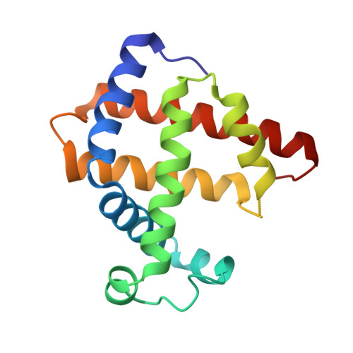
    apo-Myoglobin (apo-Mb) was reconstituted with three copper complexes: CuII(Sal-Phe) (1; Sal-Phe = N-salicylidene-L-phenylalanato), CuII(Sal-Leu) (2; Sal-Leu = N-salicylidene-L-leucinato), and CuII(Sal-Ala) (3; Sal-Ala = N-salicylidene-L-alanato). The crystal structures of 1.apo-Mb (1.65 Angstrom resolution) and 2.apo-Mb (1.8 Angstrom resolution) show that the coordination geometry around the CuII atom in apo-Mb is distorted square-planar with tridentate Sal-X and a Nepsilon atom of His64 in the apo-Mb cavity and the plane of these copper complexes is perpendicular to that of heme. These results suggest that the apo-Mb cavity can hold metal complexes with various coordination geometries.


  • Organizational Affiliation

    Department of Chemistry, Graduate School of Science, Nagoya University, Nagoya 464-8602, Japan.


Macromolecules
Find similar proteins by:  (by identity cutoff)  |  3D Structure
Entity ID: 1
MoleculeChains Sequence LengthOrganismDetailsImage
Myoglobin154Physeter catodonMutation(s): 0 
UniProt
Find proteins for P02185 (Physeter macrocephalus)
Explore P02185 
Go to UniProtKB:  P02185
Entity Groups  
Sequence Clusters30% Identity50% Identity70% Identity90% Identity95% Identity100% Identity
UniProt GroupP02185
Sequence Annotations
Expand
  • Reference Sequence
Experimental Data & Validation

Experimental Data

  • Method: X-RAY DIFFRACTION
  • Resolution: 1.80 Å
  • R-Value Free: 0.239 
  • R-Value Work: 0.189 
  • R-Value Observed: 0.192 
  • Space Group: P 21 21 21
Unit Cell:
Length ( Å )Angle ( ˚ )
a = 32.58α = 90
b = 57.901β = 90
c = 75.282γ = 90
Software Package:
Software NamePurpose
REFMACrefinement
DENZOdata reduction
SCALEPACKdata scaling
MOLREPphasing

Structure Validation

View Full Validation Report


Ligand Structure Quality Assessment 


Entry History 

Deposition Data

Revision History  (Full details and data files)

  • Version 1.0: 2007-07-03
    Type: Initial release
  • Version 1.1: 2008-04-30
    Changes: Version format compliance
  • Version 1.2: 2011-07-13
    Changes: Non-polymer description, Version format compliance
  • Version 1.3: 2023-10-25
    Changes: Data collection, Database references, Derived calculations, Refinement description