2DVW

Structure of the Oncoprotein Gankyrin in Complex with S6 ATPase of the 26S Proteasome


Experimental Data Snapshot

  • Method: X-RAY DIFFRACTION
  • Resolution: 2.30 Å
  • R-Value Free: 0.238 
  • R-Value Work: 0.169 
  • R-Value Observed: 0.169 

Starting Model: experimental
View more details

wwPDB Validation   3D Report Full Report


This is version 1.3 of the entry. See complete history


Literature

Structure of the Oncoprotein Gankyrin in Complex with S6 ATPase of the 26S Proteasome

Nakamura, Y.Nakano, K.Umehara, T.Kimura, M.Hayashizaki, Y.Tanaka, A.Horikoshi, M.Padmanabhan, B.Yokoyama, S.

(2007) Structure 15: 179-189

  • DOI: https://doi.org/10.1016/j.str.2006.11.015
  • Primary Citation of Related Structures:  
    2DVW

  • PubMed Abstract: 

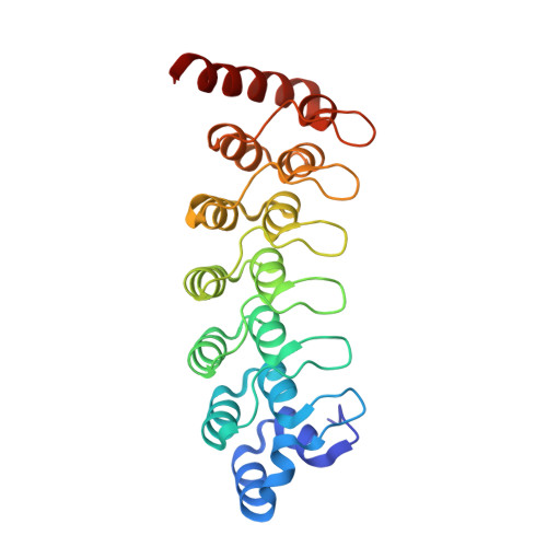
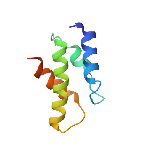
    Gankyrin is an oncoprotein commonly overexpressed in most hepatocellular carcinomas. Gankyrin interacts with S6 ATPase of the 19S regulatory particle of the 26S proteasome and enhances the degradation of the tumor suppressors pRb and p53. Here, we report the structure of gankyrin in complex with the C-terminal domain of S6 ATPase. Almost all of the seven ankyrin repeats of gankyrin interact, through its concave region, with the C-terminal domain of S6 ATPase. The intermolecular interactions occur through the complementary charged residues between gankyrin and S6 ATPase. Biochemical studies based on the structure of the complex revealed that gankyrin interacts with pRb in both the presence and absence of S6 ATPase; however, the E182 residue in gankyrin is essential for the pRb interaction. These results provide a structural basis for the involvement of gankyrin in the pRb degradation pathway, through its association with S6 ATPase of the 26S proteasome.


  • Organizational Affiliation

    RIKEN Genomic Sciences Center, 1-7-22 Suehiro-cho, Tsurumi, Yokohama 230-0045, Japan.


Macromolecules
Find similar proteins by:  (by identity cutoff)  |  3D Structure
Entity ID: 1
MoleculeChains Sequence LengthOrganismDetailsImage
26S proteasome non-ATPase regulatory subunit 10231Mus musculusMutation(s): 0 
Gene Names: Psmd10
UniProt
Find proteins for Q9Z2X2 (Mus musculus)
Explore Q9Z2X2 
Go to UniProtKB:  Q9Z2X2
Entity Groups  
Sequence Clusters30% Identity50% Identity70% Identity90% Identity95% Identity100% Identity
UniProt GroupQ9Z2X2
Sequence Annotations
Expand
  • Reference Sequence
Find similar proteins by:  (by identity cutoff)  |  3D Structure
Entity ID: 2
MoleculeChains Sequence LengthOrganismDetailsImage
26S protease regulatory subunit 6B83Homo sapiensMutation(s): 0 
Gene Names: PSMC4TBP7
UniProt & NIH Common Fund Data Resources
Find proteins for P43686 (Homo sapiens)
Explore P43686 
Go to UniProtKB:  P43686
PHAROS:  P43686
GTEx:  ENSG00000013275 
Entity Groups  
Sequence Clusters30% Identity50% Identity70% Identity90% Identity95% Identity100% Identity
UniProt GroupP43686
Sequence Annotations
Expand
  • Reference Sequence
Experimental Data & Validation

Experimental Data

  • Method: X-RAY DIFFRACTION
  • Resolution: 2.30 Å
  • R-Value Free: 0.238 
  • R-Value Work: 0.169 
  • R-Value Observed: 0.169 
  • Space Group: P 21 21 21
Unit Cell:
Length ( Å )Angle ( ˚ )
a = 45.398α = 90
b = 67.886β = 90
c = 107.767γ = 90
Software Package:
Software NamePurpose
MOLREPphasing
CNSrefinement
HKL-2000data reduction
SCALEPACKdata scaling

Structure Validation

View Full Validation Report


Entry History 

Deposition Data

Revision History  (Full details and data files)

  • Version 1.0: 2007-03-13
    Type: Initial release
  • Version 1.1: 2008-04-30
    Changes: Version format compliance
  • Version 1.2: 2011-07-13
    Changes: Version format compliance
  • Version 1.3: 2023-10-25
    Changes: Data collection, Database references, Refinement description