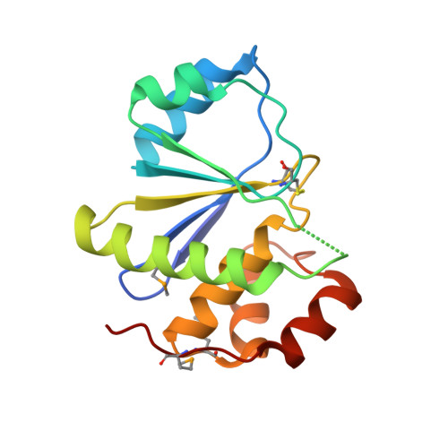
Structure and Biochemical Properties of PRL-1, a Phosphatase Implicated in Cell Growth, Differentiation, and Tumor Invasion.
Sun, J.P., Wang, W.Q., Yang, H., Liu, S., Liang, F., Fedorov, A.A., Almo, S.C., Zhang, Z.Y.(2005) Biochemistry 44: 12009-12021
- PubMed: 16142898
- DOI: https://doi.org/10.1021/bi0509191
- Primary Citation of Related Structures:
1X24, 1ZCK, 1ZCL - PubMed Abstract:
The PRL (phosphatase of regenerating liver) phosphatases constitute a novel class of small, prenylated phosphatases that are implicated in promoting cell growth, differentiation, and tumor invasion, and represent attractive targets for anticancer therapy. Here we describe the crystal structures of native PRL-1 as well as the catalytically inactive mutant PRL-1/C104S in complex with sulfate. PRL-1 exists as a trimer in the crystalline state, burying 1140 A2 of accessible surface area at each dimer interface. Trimerization creates a large, bipartite membrane-binding surface in which the exposed C-terminal basic residues could cooperate with the adjacent prenylation group to anchor PRL-1 on the acidic inner membrane. Structural and kinetic analyses place PRL-1 in the family of dual specificity phopsphatases with closest structural similarity to the Cdc14 phosphatase and provide a molecular basis for catalytic activation of the PRL phosphatases. Finally, native PRL-1 is crystallized in an oxidized form in which a disulfide is formed between the active site Cys104 and a neighboring residue Cys49, which blocks both substrate binding and catalysis. Biochemical studies in solution and in the cell support a potential regulatory role of this intramolecular disulfide bond formation in response to reactive oxygen species such as H2O2.
Organizational Affiliation:
Department of Molecular Pharmacology, Albert Einstein College of Medicine, 1300 Morris Park Avenue, Bronx, New York 10461, USA.


