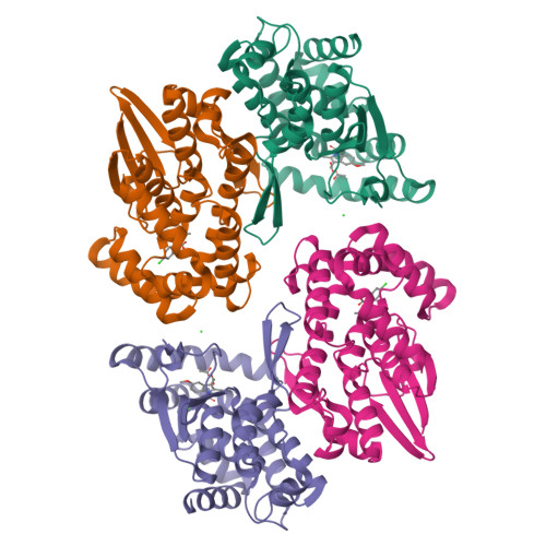
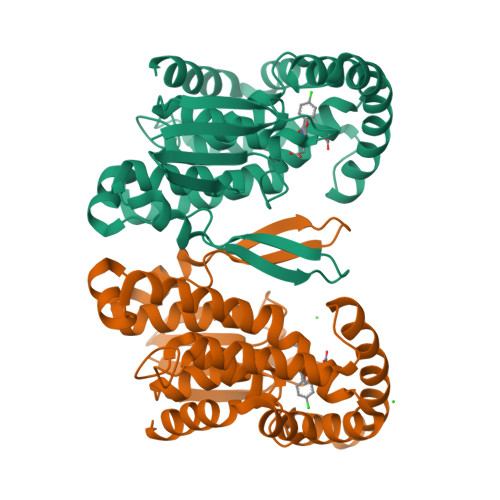
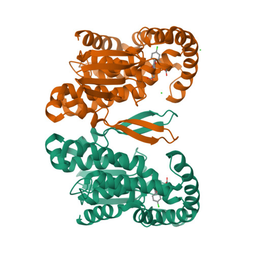
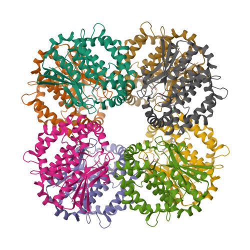
1Z9H

Microsomal prostaglandin E synthase type-2


Experimental Data Snapshot

  • Method: X-RAY DIFFRACTION
  • Resolution: 2.60 Å
  • R-Value Free: 0.251 
  • R-Value Work: 0.214 
  • R-Value Observed: 0.215 

wwPDB Validation   3D Report Full Report


Ligand Structure Quality Assessment 


This is version 1.4 of the entry. See complete history


Literature

Crystal Structure and Possible Catalytic Mechanism of Microsomal Prostaglandin E Synthase Type 2 (mPGES-2).

Yamada, T.Komoto, J.Watanabe, K.Ohmiya, Y.Takusagawa, F.

(2005) J Mol Biol 348: 1163-1176

  • DOI: https://doi.org/10.1016/j.jmb.2005.03.035
  • Primary Citation of Related Structures:  
    1Z9H

  • PubMed Abstract: 

    Prostaglandin (PG) H(2) (PGH(2)), formed from arachidonic acid, is an unstable intermediate and is converted efficiently into more stable arachidonate metabolites (PGD(2), PGE(2), and PGF(2)) by the action of three groups of enzymes. Prostaglandin E synthase catalyzes an isomerization reaction, PGH(2) to PGE(2). Microsomal prostaglandin E synthase type-2 (mPGES-2) has been crystallized with an anti-inflammatory drug indomethacin (IMN), and the complex structure has been determined at 2.6A resolution. mPGES-2 forms a dimer and is attached to lipid membrane by anchoring the N-terminal section. Two hydrophobic pockets connected to form a V shape are located in the bottom of a large cavity. IMN binds deeply in the cavity by placing the OMe-indole and chlorophenyl moieties into the V-shaped pockets, respectively, and the carboxyl group interacts with S(gamma) of C110 by forming a H-bond. A characteristic H-bond chain formation (N-H...S(gamma)-H...S(gamma)...H-N) is seen through Y107-C113-C110-F112, which apparently decreases the pK(a) of S(gamma) of C110. The geometry suggests that the S(gamma) of C110 is most likely the catalytic site of mPGES-2. A search of the RCSB Protein Data Bank suggests that IMN can fit into the PGH(2) binding site in various proteins. On the basis of the crystal structure and mutation data, a PGH(2)-bound model structure was built. PGH(2) fits well into the IMN binding site by placing the alpha and omega-chains in the V-shaped pockets, and the endoperoxide moiety interacts with S(gamma) of C110. A possible catalytic mechanism is proposed on the basis of the crystal and model structures, and an alternative catalytic mechanism is described. The fold of mPGES-2 is quite similar to those of GSH-dependent hematopoietic prostaglandin D synthase, except for the two large loop sections.


  • Organizational Affiliation

    Department of Molecular Biosciences, University of Kansas, 1200 Sunnyside Ave, Lawrence, KS 66045-7534, USA.


Macromolecules
Find similar proteins by:  (by identity cutoff)  |  3D Structure
Entity ID: 1
MoleculeChains Sequence LengthOrganismDetailsImage
membrane-associated prostaglandin E synthase-2
A, B, C, D
290Macaca fascicularisMutation(s): 0 
EC: 5.3.99.3
Membrane Entity: Yes 
UniProt
Find proteins for Q9N0A4 (Macaca fascicularis)
Explore Q9N0A4 
Go to UniProtKB:  Q9N0A4
Entity Groups  
Sequence Clusters30% Identity50% Identity70% Identity90% Identity95% Identity100% Identity
UniProt GroupQ9N0A4
Sequence Annotations
Expand
  • Reference Sequence
Small Molecules
Ligands 3 Unique
IDChains Name / Formula / InChI Key2D Diagram3D Interactions
IMN
Query on IMN

Download Ideal Coordinates CCD File 
F [auth A],
J [auth B],
L [auth C],
P [auth D]
INDOMETHACIN
C19 H16 Cl N O4
CGIGDMFJXJATDK-UHFFFAOYSA-N
ACT
Query on ACT

Download Ideal Coordinates CCD File 
E [auth A],
H [auth B],
K [auth C],
O [auth D]
ACETATE ION
C2 H3 O2
QTBSBXVTEAMEQO-UHFFFAOYSA-M
CL
Query on CL

Download Ideal Coordinates CCD File 
G [auth B],
I [auth B],
M [auth D],
N [auth D]
CHLORIDE ION
Cl
VEXZGXHMUGYJMC-UHFFFAOYSA-M
Binding Affinity Annotations 
IDSourceBinding Affinity
IMN PDBBind:  1Z9H IC50: 1.00e+6 (nM) from 1 assay(s)
Experimental Data & Validation

Experimental Data

  • Method: X-RAY DIFFRACTION
  • Resolution: 2.60 Å
  • R-Value Free: 0.251 
  • R-Value Work: 0.214 
  • R-Value Observed: 0.215 
  • Space Group: C 1 2 1
Unit Cell:
Length ( Å )Angle ( ˚ )
a = 128.24α = 90
b = 122.83β = 110.6
c = 111.53γ = 90
Software Package:
Software NamePurpose
APSdata collection
SCALEPACKdata scaling
CNSrefinement
CNSphasing

Structure Validation

View Full Validation Report


Ligand Structure Quality Assessment 


Entry History 

Deposition Data

Revision History  (Full details and data files)

  • Version 1.0: 2005-05-17
    Type: Initial release
  • Version 1.1: 2008-04-30
    Changes: Version format compliance
  • Version 1.2: 2011-07-13
    Changes: Version format compliance
  • Version 1.3: 2017-10-11
    Changes: Refinement description
  • Version 1.4: 2024-02-14
    Changes: Data collection, Database references, Derived calculations