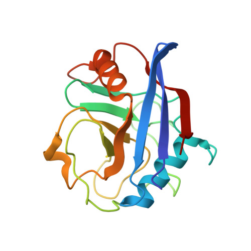
1YND

Structure of human cyclophilin A in complex with the novel immunosuppressant sanglifehrin A at 1.6A resolution


Experimental Data Snapshot

  • Method: X-RAY DIFFRACTION
  • Resolution: 1.60 Å
  • R-Value Free: 0.185 
  • R-Value Work: 0.163 
  • R-Value Observed: 0.164 

Starting Model: experimental
View more details

wwPDB Validation   3D Report Full Report


Ligand Structure Quality Assessment 


This is version 1.3 of the entry. See complete history


Literature

Structure of human cyclophilin A in complex with the novel immunosuppressant sanglifehrin A at 1.6 A resolution.

Kallen, J.Sedrani, R.Zenke, G.Wagner, J.

(2005) J Biol Chem 280: 21965-21971

  • DOI: https://doi.org/10.1074/jbc.M501623200
  • Primary Citation of Related Structures:  
    1YND

  • PubMed Abstract: 

    Sanglifehrin A (SFA) is a novel immunosuppressant isolated from Streptomyces sp. that binds strongly to the human immunophilin cyclophilin A (CypA). SFA exerts its immunosuppressive activity through a mode of action different from that of all other known immunophilin-binding substances, namely cyclosporine A (CsA), FK506, and rapamycin. We have determined the crystal structure of human CypA in complex with SFA at 1.6 A resolution. The high resolution of the structure revealed the absolute configuration at all 17 chiral centers of SFA as well as the details of the CypA/SFA interactions. In particular, it was shown that the 22-membered macrocycle of SFA is deeply embedded in the same binding site as CsA and forms six direct hydrogen bonds with CypA. The effector domain of SFA, on the other hand, has a chemical and three-dimensional structure very different from CsA, already strongly suggesting different immunosuppressive mechanisms. Furthermore, two CypA.SFA complexes form a dimer in the crystal as well as in solution as shown by light scattering and size exclusion chromatography experiments. This observation raises the possibility that the dimer of CypA.SFA complexes is the molecular species mediating the immunosuppressive effect.


  • Organizational Affiliation

    Protein Structure Unit, Novartis Institutes for BioMedical Research, CH-4002 Basel, Switzerland. joerg.kallen@pharma.novartis.com


Macromolecules
Find similar proteins by:  (by identity cutoff)  |  3D Structure
Entity ID: 1
MoleculeChains Sequence LengthOrganismDetailsImage
Peptidyl-prolyl cis-trans isomerase A
A, B
165Homo sapiensMutation(s): 0 
Gene Names: PPIACYPA
EC: 5.2.1.8
UniProt & NIH Common Fund Data Resources
Find proteins for P62937 (Homo sapiens)
Explore P62937 
Go to UniProtKB:  P62937
PHAROS:  P62937
GTEx:  ENSG00000196262 
Entity Groups  
Sequence Clusters30% Identity50% Identity70% Identity90% Identity95% Identity100% Identity
UniProt GroupP62937
Sequence Annotations
Expand
  • Reference Sequence
Small Molecules
Binding Affinity Annotations 
IDSourceBinding Affinity
SFA BindingDB:  1YND Kd: 3.3 (nM) from 1 assay(s)
PDBBind:  1YND IC50: 3.6 (nM) from 1 assay(s)
Experimental Data & Validation

Experimental Data

  • Method: X-RAY DIFFRACTION
  • Resolution: 1.60 Å
  • R-Value Free: 0.185 
  • R-Value Work: 0.163 
  • R-Value Observed: 0.164 
  • Space Group: P 21 21 21
Unit Cell:
Length ( Å )Angle ( ˚ )
a = 62.6α = 90
b = 65.4β = 90
c = 76.6γ = 90
Software Package:
Software NamePurpose
REFMACrefinement
DENZOdata reduction
SCALEPACKdata scaling
X-PLORphasing

Structure Validation

View Full Validation Report


Ligand Structure Quality Assessment 


Entry History 

Deposition Data

Revision History  (Full details and data files)

  • Version 1.0: 2005-04-05
    Type: Initial release
  • Version 1.1: 2008-04-30
    Changes: Version format compliance
  • Version 1.2: 2011-07-13
    Changes: Version format compliance
  • Version 1.3: 2023-08-23
    Changes: Data collection, Database references, Derived calculations, Refinement description, Structure summary