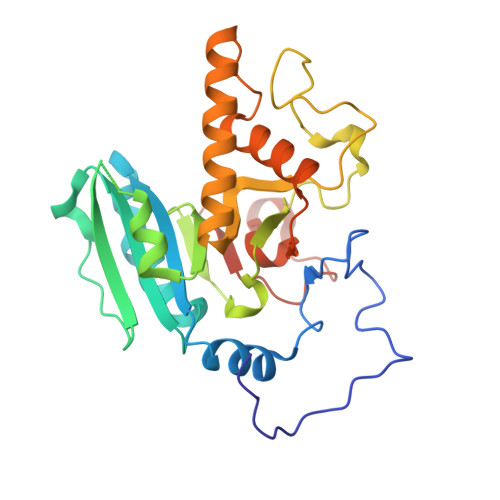
1XZ6

Mutant ABO(H) blood group glycosyltransferase A


Experimental Data Snapshot

  • Method: X-RAY DIFFRACTION
  • Resolution: 1.55 Å
  • R-Value Free: 0.243 
  • R-Value Work: 0.209 
  • R-Value Observed: 0.222 

wwPDB Validation   3D Report Full Report


This is version 1.7 of the entry. See complete history


Literature

Structural basis for the inactivity of human blood group O2 glycosyltransferase

Lee, H.J.Barry, C.H.Borisova, S.N.Seto, N.O.L.Zheng, R.B.Blancher, A.Evans, S.V.Palcic, M.M.

(2005) J Biol Chem 280: 525-529

  • DOI: https://doi.org/10.1074/jbc.M410245200
  • Primary Citation of Related Structures:  
    1WSZ, 1WT0, 1WT1, 1WT2, 1WT3, 1XZ6

  • PubMed Abstract: 

    The human ABO(H) blood group antigens are carbohydrate structures generated by glycosyltransferase enzymes. Glycosyltransferase A (GTA) uses UDP-GalNAc as a donor to transfer a monosaccharide residue to Fuc alpha1-2Gal beta-R (H)-terminating acceptors. Similarly, glycosyltransferase B (GTB) catalyzes the transfer of a monosaccharide residue from UDP-Gal to the same acceptors. These are highly homologous enzymes differing in only four of 354 amino acids, Arg/Gly-176, Gly/Ser-235, Leu/Met-266, and Gly/Ala-268. Blood group O usually stems from the expression of truncated inactive forms of GTA or GTB. Recently, an O(2) enzyme was discovered that was a full-length form of GTA with three mutations, P74S, R176G, and G268R. We showed previously that the R176G mutation increased catalytic activity with minor effects on substrate binding. Enzyme kinetics and high resolution structural studies of mutant enzymes based on the O(2) blood group transferase reveal that whereas the P74S mutation in the stem region of the protein does not appear to play a role in enzyme inactivation, the G268R mutation completely blocks the donor GalNAc-binding site leaving the acceptor binding site unaffected.


  • Organizational Affiliation

    Department of Chemistry, University of Alberta, Edmonton, Alberta T6G 2G2, Canada.


Macromolecules
Find similar proteins by:  (by identity cutoff)  |  3D Structure
Entity ID: 1
MoleculeChains Sequence LengthOrganismDetailsImage
Histo-blood group ABO system transferase292Homo sapiensMutation(s): 1 
Gene Names: ABO
EC: 2.4.1.40 (PDB Primary Data), 2.4.1.37 (UniProt)
UniProt & NIH Common Fund Data Resources
Find proteins for P16442 (Homo sapiens)
Explore P16442 
Go to UniProtKB:  P16442
PHAROS:  P16442
Entity Groups  
Sequence Clusters30% Identity50% Identity70% Identity90% Identity95% Identity100% Identity
UniProt GroupP16442
Sequence Annotations
Expand
  • Reference Sequence
Experimental Data & Validation

Experimental Data

  • Method: X-RAY DIFFRACTION
  • Resolution: 1.55 Å
  • R-Value Free: 0.243 
  • R-Value Work: 0.209 
  • R-Value Observed: 0.222 
  • Space Group: C 2 2 21
Unit Cell:
Length ( Å )Angle ( ˚ )
a = 52.9α = 90
b = 150.9β = 90
c = 79.6γ = 90
Software Package:
Software NamePurpose
CNSrefinement
CNSphasing

Structure Validation

View Full Validation Report


Entry History 

Deposition Data

Revision History  (Full details and data files)

  • Version 1.0: 2004-12-07
    Type: Initial release
  • Version 1.1: 2008-04-30
    Changes: Version format compliance
  • Version 1.2: 2011-07-13
    Changes: Derived calculations, Version format compliance
  • Version 1.3: 2017-10-11
    Changes: Refinement description
  • Version 1.4: 2018-01-31
    Changes: Experimental preparation
  • Version 1.5: 2018-03-07
    Changes: Data collection
  • Version 1.6: 2021-10-20
    Changes: Database references, Derived calculations
  • Version 1.7: 2024-02-14
    Changes: Data collection