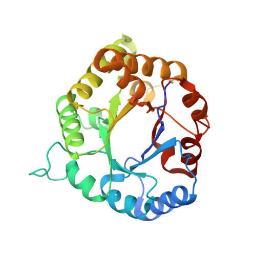
1WYI

human triosephosphate isomerase of new crystal form


Experimental Data Snapshot

  • Method: X-RAY DIFFRACTION
  • Resolution: 2.20 Å
  • R-Value Free: 0.296 
  • R-Value Work: 0.247 
  • R-Value Observed: 0.266 

wwPDB Validation   3D Report Full Report


This is version 1.4 of the entry. See complete history


Literature

Structure of a high-resolution crystal form of human triosephosphate isomerase: improvement of crystals using the gel-tube method.

Kinoshita, T.Maruki, R.Warizaya, M.Nakajima, H.Nishimura, S.

(2005) Acta Crystallogr Sect F Struct Biol Cryst Commun 61: 346-349

  • DOI: https://doi.org/10.1107/S1744309105008341
  • Primary Citation of Related Structures:  
    1WYI

  • PubMed Abstract: 

    Crystals of human triosephosphate isomerase with two crystal morphologies were obtained using the normal vapour-diffusion technique with identical crystallization conditions. One had a disordered plate shape and the crystals were hollow (crystal form 1). As a result, this form was very fragile, diffracted to 2.8 A resolution and had similar crystallographic parameters to those of the structure 1hti in the Protein Data Bank. The other had a fine needle shape (crystal form 2) and was formed more abundantly than crystal form 1, but was unsuitable for structure analysis. Since the normal vapour-diffusion method could not control the crystal morphology, gel-tube methods, both on earth and under microgravity, were applied for crystallization in order to control and improve the crystal quality. Whereas crystal form 1 was only slightly improved using this method, crystal form 2 was greatly improved and diffracted to 2.2 A resolution. Crystal form 2 contained a homodimer in the asymmetric unit, which was biologically essential. Its overall structure was similar to that of 1hti except for the flexible loop, which was located at the active centre Lys13.


  • Organizational Affiliation

    Exploratory Research Laboratories, Fujisawa Pharmaceutical Co. Ltd, 5-2-3 Tokodai, Tsukuba, Ibaraki 300-2698, Japan. takayoshi_kinoshita@po.fujisawa.co.jp


Macromolecules
Find similar proteins by:  (by identity cutoff)  |  3D Structure
Entity ID: 1
MoleculeChains Sequence LengthOrganismDetailsImage
Triosephosphate isomerase
A, B
250Homo sapiensMutation(s): 0 
EC: 5.3.1.1 (PDB Primary Data), 4.2.3.3 (UniProt)
UniProt & NIH Common Fund Data Resources
Find proteins for P60174 (Homo sapiens)
Explore P60174 
Go to UniProtKB:  P60174
PHAROS:  P60174
GTEx:  ENSG00000111669 
Entity Groups  
Sequence Clusters30% Identity50% Identity70% Identity90% Identity95% Identity100% Identity
UniProt GroupP60174
Sequence Annotations
Expand
  • Reference Sequence
Experimental Data & Validation

Experimental Data

  • Method: X-RAY DIFFRACTION
  • Resolution: 2.20 Å
  • R-Value Free: 0.296 
  • R-Value Work: 0.247 
  • R-Value Observed: 0.266 
  • Space Group: P 21 21 21
Unit Cell:
Length ( Å )Angle ( ˚ )
a = 46.564α = 90
b = 70.883β = 90
c = 141.412γ = 90
Software Package:
Software NamePurpose
CNXrefinement
CrystalCleardata scaling
CNXphasing

Structure Validation

View Full Validation Report


Entry History 

Deposition Data

Revision History  (Full details and data files)

  • Version 1.0: 2005-04-12
    Type: Initial release
  • Version 1.1: 2008-04-30
    Changes: Version format compliance
  • Version 1.2: 2011-07-13
    Changes: Derived calculations, Version format compliance
  • Version 1.3: 2017-10-11
    Changes: Refinement description
  • Version 1.4: 2024-03-13
    Changes: Data collection, Database references