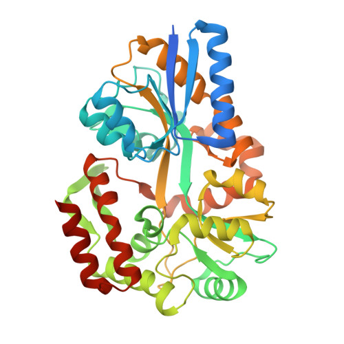
1URS

X-ray structures of the maltose-maltodextrin binding protein of the thermoacidophilic bacterium Alicyclobacillus acidocaldarius


Experimental Data Snapshot

  • Method: X-RAY DIFFRACTION
  • Resolution: 1.45 Å
  • R-Value Free: 0.232 
  • R-Value Work: 0.210 
  • R-Value Observed: 0.210 

Starting Model: experimental
View more details

wwPDB Validation   3D Report Full Report


This is version 2.1 of the entry. See complete history


Literature

X-Ray Structures of the Maltose-Maltodextrin-Binding Protein of the Thermoacidophilic Bacterium Alicyclobacillus Acidocaldarius Provide Insight Into Acid Stability of Proteins

Schafer, K.Magnusson, U.Scheffel, F.Schiefner, A.Sandgren, M.O.J.Diederichs, K.Welte, W.Hulsmann, A.Schneider, E.Mowbray, S.L.

(2004) J Mol Biol 335: 261

  • DOI: https://doi.org/10.1016/j.jmb.2003.10.042
  • Primary Citation of Related Structures:  
    1URD, 1URG, 1URS

  • PubMed Abstract: 

    Maltose-binding proteins act as primary receptors in bacterial transport and chemotaxis systems. We report here crystal structures of the thermoacidostable maltose-binding protein from Alicyclobacillus acidocaldarius, and explore its modes of binding to maltose and maltotriose. Further, comparison with the structures of related proteins from Escherichia coli (a mesophile), and two hyperthermophiles (Pyrococcus furiosus and Thermococcus litoralis) allows an investigation of the basis of thermo- and acidostability in this family of proteins.The thermoacidophilic protein has fewer charged residues than the other three structures, which is compensated by an increase in the number of polar residues. Although the content of acidic and basic residues is approximately equal, more basic residues are exposed on its surface whereas most acidic residues are buried in the interior. As a consequence, this protein has a highly positive surface charge. Fewer salt bridges are buried than in the other MBP structures, but the number exposed on its surface does not appear to be unusual. These features appear to be correlated with the acidostability of the A. acidocaldarius protein rather than its thermostability. An analysis of cavities within the proteins shows that the extremophile proteins are more closely packed than the mesophilic one. Proline content is slightly higher in the hyperthermophiles and thermoacidophiles than in mesophiles, and this amino acid is more common at the second position of beta-turns, properties that are also probably related to thermostability. Secondary structural content does not vary greatly in the different structures, and so is not a contributing factor.


  • Organizational Affiliation

    Fachbereich Biologie, Universität Konstanz, Universitätsstrasse 10, 78457, Konstanz, Germany.


Macromolecules
Find similar proteins by:  (by identity cutoff)  |  3D Structure
Entity ID: 1
MoleculeChains Sequence LengthOrganismDetailsImage
MALTOSE-BINDING PROTEIN
A, B
402Alicyclobacillus acidocaldariusMutation(s): 0 
UniProt
Find proteins for Q9RHZ6 (Alicyclobacillus acidocaldarius subsp. acidocaldarius)
Explore Q9RHZ6 
Go to UniProtKB:  Q9RHZ6
Entity Groups  
Sequence Clusters30% Identity50% Identity70% Identity90% Identity95% Identity100% Identity
UniProt GroupQ9RHZ6
Sequence Annotations
Expand
  • Reference Sequence
Oligosaccharides

Help

Entity ID: 2
MoleculeChains Length2D Diagram Glycosylation3D Interactions
alpha-D-glucopyranose-(1-4)-alpha-D-glucopyranose-(1-4)-alpha-D-glucopyranose
C, D
3N/A
Glycosylation Resources
GlyTouCan:  G96370VA
GlyCosmos:  G96370VA
GlyGen:  G96370VA
Biologically Interesting Molecules (External Reference) 1 Unique
Experimental Data & Validation

Experimental Data

  • Method: X-RAY DIFFRACTION
  • Resolution: 1.45 Å
  • R-Value Free: 0.232 
  • R-Value Work: 0.210 
  • R-Value Observed: 0.210 
  • Space Group: P 1 21 1
Unit Cell:
Length ( Å )Angle ( ˚ )
a = 49.18α = 90
b = 70.53β = 96.98
c = 104.06γ = 90
Software Package:
Software NamePurpose
CNSrefinement
XDSdata reduction
XDSdata scaling
AMoREphasing

Structure Validation

View Full Validation Report


Entry History 

Deposition Data

Revision History  (Full details and data files)

  • Version 1.0: 2003-12-11
    Type: Initial release
  • Version 1.1: 2011-05-08
    Changes: Version format compliance
  • Version 1.2: 2011-07-13
    Changes: Version format compliance
  • Version 2.0: 2020-07-29
    Type: Remediation
    Reason: Carbohydrate remediation
    Changes: Atomic model, Data collection, Derived calculations, Non-polymer description, Other, Structure summary
  • Version 2.1: 2023-12-13
    Changes: Data collection, Database references, Refinement description, Structure summary