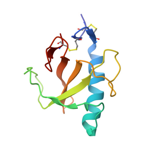
1TTO

Crystal structure of the Rnase T1 variant R2


Experimental Data Snapshot

  • Method: X-RAY DIFFRACTION
  • Resolution: 2.10 Å
  • R-Value Free: 0.238 
  • R-Value Work: 0.187 

Starting Model: experimental
View more details

wwPDB Validation   3D Report Full Report


This is version 1.4 of the entry. See complete history


Literature

Purine activity of RNase T1RV is further improved by substitution of Trp59 by tyrosine

Czaja, R.Perbandt, M.Betzel, C.Hahn, U.

(2005) Biochem Biophys Res Commun 336: 882-889

  • DOI: https://doi.org/10.1016/j.bbrc.2005.08.188
  • Primary Citation of Related Structures:  
    1TTO

  • PubMed Abstract: 

    Ribonuclease T1 is an enzyme that cleaves single-stranded RNA with high specificity after guanylyl residues. Although this enzyme is a very good characterized protein with respect to structure and enzymatic function, we were only recently successful in generating RNase T1-RV, a variant where the specificity was changed from guanine to purine. As this change of substrate specificity was made at the cost of activity, the aim was now to further improve the overall activity of the enzyme. Therefore, we have substituted the tryptophan in position 59 by tyrosine. This substitution led to an increase of enzymatic activity in comparison to variant RV to 425%. As the extent of this enhancement is unique so far we have crystallized and analyzed the structure of this variant in order to get more insights into the reasons for this. Here, we present the crystal structure of this so-called RNase T1-R2 at 2.1A resolution. The structure was determined by molecular replacement using the coordinates of the RV variant (PDB entry: 1Q9E). The data were refined to an R-factor of 18.7% and R(free) of 24%, respectively. The asymmetric unit contains three molecules and the crystal packing is very similar to that of variant RV.


  • Organizational Affiliation

    Department of Chemistry, Division of Biochemistry and Molecular Biology, University of Hamburg, Martin-Luther-King-Platz 6, 20146 Hamburg, Germany.


Macromolecules
Find similar proteins by:  (by identity cutoff)  |  3D Structure
Entity ID: 1
MoleculeChains Sequence LengthOrganismDetailsImage
RNase T1
A, B, C
104Aspergillus oryzaeMutation(s): 6 
EC: 3.1.27.3 (PDB Primary Data), 4.6.1.24 (UniProt)
UniProt
Find proteins for P00651 (Aspergillus oryzae (strain ATCC 42149 / RIB 40))
Explore P00651 
Go to UniProtKB:  P00651
Entity Groups  
Sequence Clusters30% Identity50% Identity70% Identity90% Identity95% Identity100% Identity
UniProt GroupP00651
Sequence Annotations
Expand
  • Reference Sequence
Experimental Data & Validation

Experimental Data

  • Method: X-RAY DIFFRACTION
  • Resolution: 2.10 Å
  • R-Value Free: 0.238 
  • R-Value Work: 0.187 
  • Space Group: P 61
Unit Cell:
Length ( Å )Angle ( ˚ )
a = 56.524α = 90
b = 56.524β = 90
c = 159.21γ = 120
Software Package:
Software NamePurpose
DENZOdata reduction
SCALEPACKdata scaling
AMoREphasing
CNSrefinement

Structure Validation

View Full Validation Report


Entry History 

Deposition Data

Revision History  (Full details and data files)

  • Version 1.0: 2005-09-06
    Type: Initial release
  • Version 1.1: 2008-04-30
    Changes: Version format compliance
  • Version 1.2: 2011-07-13
    Changes: Version format compliance
  • Version 1.3: 2021-11-10
    Changes: Data collection, Database references, Derived calculations
  • Version 1.4: 2023-10-25
    Changes: Data collection, Refinement description