1THJ

CARBONIC ANHYDRASE FROM METHANOSARCINA


Experimental Data Snapshot

  • Method: X-RAY DIFFRACTION
  • Resolution: 2.80 Å
  • R-Value Free: 0.295 
  • R-Value Work: 0.234 
  • R-Value Observed: 0.234 

wwPDB Validation   3D Report Full Report


This is version 1.3 of the entry. See complete history


Literature

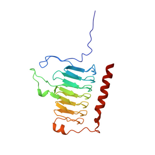
A left-hand beta-helix revealed by the crystal structure of a carbonic anhydrase from the archaeon Methanosarcina thermophila.

Kisker, C.Schindelin, H.Alber, B.E.Ferry, J.G.Rees, D.C.

(1996) EMBO J 15: 2323-2330

  • Primary Citation of Related Structures:  
    1THJ

  • PubMed Abstract: 

    A carbonic anhydrase from the thermophilic archaeon Methanosarcina thermophila that exhibits no significant sequence similarity to known carbonic anhydrases has recently been characterized. Here we present the structure of this enzyme, which adopts a left-handed parallel beta-helix fold. This fold is of particular interest since it contains only left-handed crossover connections between the parallel beta-strands, which so far have been observed very infrequently. The active form of the enzyme is a trimer with three zinc-containing active sites, each located at the interface between two monomers. While the arrangement of active site groups differs between this enzyme and the carbonic anhydrases from higher vertebrates, there are structural similarities in the zinc coordination environment, suggestive of convergent evolution dictated by the chemical requirements for catalysis of the same reaction. Based on sequence similarities, the structure of this enzyme is the prototype of a new class of carbonic anhydrases with representatives in all three phylogenetic domains of life.


  • Organizational Affiliation

    Division of Chemistry and Chemical Engineering, California Institute of Technology, Pasadena, CA 91125, USA. kisker@citray.caltech.edu


Macromolecules
Find similar proteins by:  (by identity cutoff)  |  3D Structure
Entity ID: 1
MoleculeChains Sequence LengthOrganismDetailsImage
CARBONIC ANHYDRASE
A, B, C
214Methanosarcina thermophilaMutation(s): 0 
EC: 4.2.1.1
UniProt
Find proteins for P40881 (Methanosarcina thermophila (strain ATCC 43570 / DSM 1825 / OCM 12 / VKM B-1830 / TM-1))
Explore P40881 
Go to UniProtKB:  P40881
Entity Groups  
Sequence Clusters30% Identity50% Identity70% Identity90% Identity95% Identity100% Identity
UniProt GroupP40881
Sequence Annotations
Expand
  • Reference Sequence
Experimental Data & Validation

Experimental Data

  • Method: X-RAY DIFFRACTION
  • Resolution: 2.80 Å
  • R-Value Free: 0.295 
  • R-Value Work: 0.234 
  • R-Value Observed: 0.234 
  • Space Group: P 43 21 2
Unit Cell:
Length ( Å )Angle ( ˚ )
a = 71.84α = 90
b = 71.84β = 90
c = 333.513γ = 90
Software Package:
Software NamePurpose
X-PLORmodel building
X-PLORrefinement
DENZOdata reduction
Agrovatadata scaling
ROTAVATAdata scaling
X-PLORphasing

Structure Validation

View Full Validation Report


Entry History 

Deposition Data

Revision History  (Full details and data files)

  • Version 1.0: 1996-10-14
    Type: Initial release
  • Version 1.1: 2008-03-24
    Changes: Version format compliance
  • Version 1.2: 2011-07-13
    Changes: Version format compliance
  • Version 1.3: 2024-02-14
    Changes: Data collection, Database references, Derived calculations, Other, Refinement description