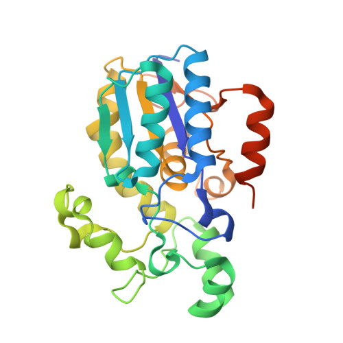
Crystal structure of human bisphosphoglycerate mutase
Wang, Y., Wei, Z., Bian, Q., Cheng, Z., Wan, M., Liu, L., Gong, W.(2004) J Biol Chem 279: 39132-39138
- PubMed: 15258155
- DOI: https://doi.org/10.1074/jbc.M405982200
- Primary Citation of Related Structures:
1T8P - PubMed Abstract:
Bisphosphoglycerate mutase is a trifunctional enzyme of which the main function is to synthesize 2,3-bisphosphoglycerate, the allosteric effector of hemoglobin. The gene coding for bisphosphoglycerate mutase from the human cDNA library was cloned and expressed in Escherichia coli. The protein crystals were obtained and diffract to 2.5 A and produced the first crystal structure of bisphosphoglycerate mutase. The model was refined to a crystallographic R-factor of 0.200 and R(free) of 0.266 with excellent stereochemistry. The enzyme remains a dimer in the crystal. The overall structure of the enzyme resembles that of the cofactor-dependent phosphoglycerate mutase except the regions of 13-21, 98-117, 127-151, and the C-terminal tail. The conformational changes in the backbone and the side chains of some residues reveal the structural basis for the different activities between phosphoglycerate mutase and bisphosphoglycerate mutase. The bisphosphoglycerate mutase-specific residue Gly-14 may cause the most important conformational changes, which makes the side chain of Glu-13 orient toward the active site. The positions of Glu-13 and Phe-22 prevent 2,3-bisphosphoglycerate from binding in the way proposed previously. In addition, the side chain of Glu-13 would affect the Glu-89 protonation ability responsible for the low mutase activity. Other structural variations, which could be connected with functional differences, are also discussed.
Organizational Affiliation:
National Laboratory of Biomacromolecules, Institute of Biophysics, Chinese Academy of Sciences, Beijing 100101, China.


