1SL8

Calcium-loaded apo-aequorin from Aequorea victoria


Experimental Data Snapshot

  • Method: X-RAY DIFFRACTION
  • Resolution: 1.70 Å
  • R-Value Free: 0.236 
  • R-Value Work: 0.216 
  • R-Value Observed: 0.217 

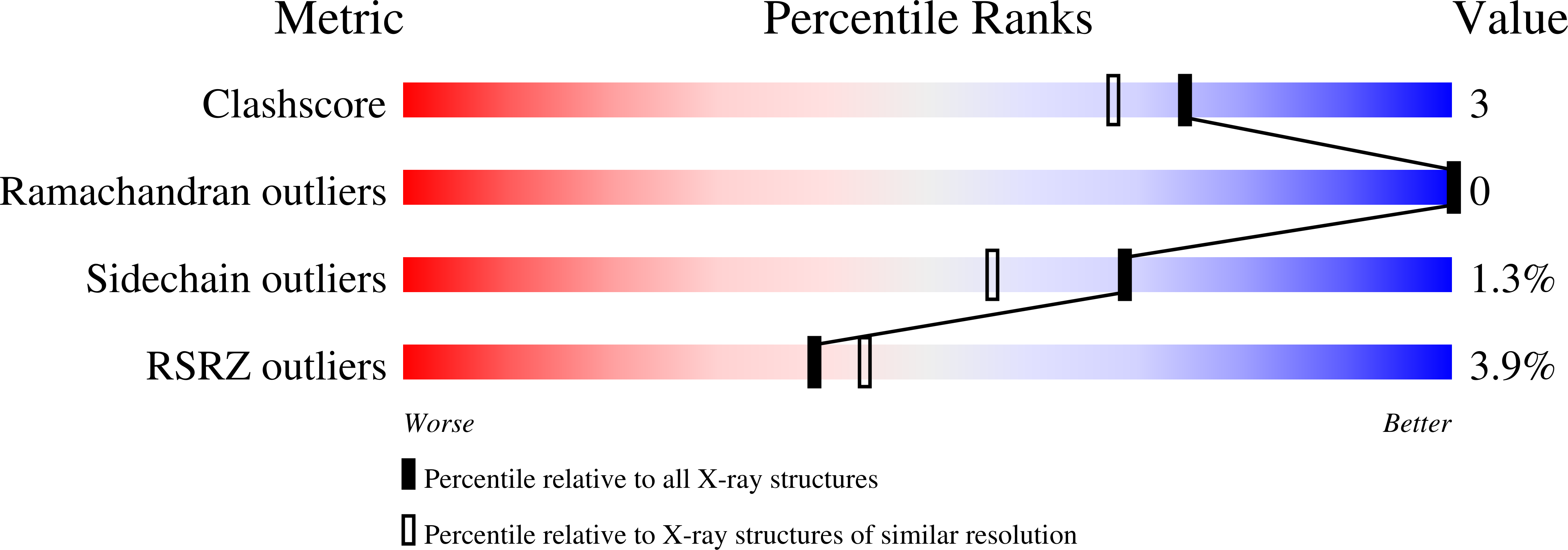
wwPDB Validation   3D Report Full Report


This is version 1.3 of the entry. See complete history


Literature

All three Ca2+-binding loops of photoproteins bind calcium ions: The crystal structures of calcium-loaded apo-aequorin and apo-obelin.

Deng, L.Vysotski, E.S.Markova, S.V.Liu, Z.J.Lee, J.Rose, J.Wang, B.C.

(2005) Protein Sci 14: 663-675

  • DOI: https://doi.org/10.1110/ps.041142905
  • Primary Citation of Related Structures:  
    1SL7, 1SL8

  • PubMed Abstract: 

    The crystal structures of calcium-loaded apo-aequorin and apo-obelin have been determined at resolutions 1.7A and 2.2 A, respectively. A calcium ion is observed in each of the three EF-hand loops that have the canonical calcium-binding sequence, and each is coordinated in the characteristic pentagonal bipyramidal configuration. The calcium-loaded apo-protein retain the same compact scaffold and overall fold as the unreacted photoproteins containing the bound substrate, 2-hyroperoxycoelenterazine, and also the same as the Ca2+-discharged obelin bound with product, coleneteramide. Nevertheless, there are easily discerned shifts in both helix and loop regions, and the shifts are not the same between the two proteins. It is suggested that these photoproteins to sense Ca2+ concentration transients and to produce their bioluminescence response on the millisecond timescale. A mechanism of intrastructural transmission of the calcium signal is proposed.


  • Organizational Affiliation

    Department of Biochemistry and Molecular Biology, University of Georgia, Athens, GA 30602, USA.


Macromolecules
Find similar proteins by:  (by identity cutoff)  |  3D Structure
Entity ID: 1
MoleculeChains Sequence LengthOrganismDetailsImage
Aequorin 1191Aequorea victoriaMutation(s): 0 
UniProt
Find proteins for P07164 (Aequorea victoria)
Explore P07164 
Go to UniProtKB:  P07164
Entity Groups  
Sequence Clusters30% Identity50% Identity70% Identity90% Identity95% Identity100% Identity
UniProt GroupP07164
Sequence Annotations
Expand
  • Reference Sequence
Experimental Data & Validation

Experimental Data

  • Method: X-RAY DIFFRACTION
  • Resolution: 1.70 Å
  • R-Value Free: 0.236 
  • R-Value Work: 0.216 
  • R-Value Observed: 0.217 
  • Space Group: P 43 21 2
Unit Cell:
Length ( Å )Angle ( ˚ )
a = 54.371α = 90
b = 54.371β = 90
c = 135.109γ = 90
Software Package:
Software NamePurpose
REFMACrefinement
HKL-2000data reduction
SCALEPACKdata scaling
SOLVEphasing

Structure Validation

View Full Validation Report


Entry History 

Revision History  (Full details and data files)

  • Version 1.0: 2004-12-28
    Type: Initial release
  • Version 1.1: 2008-04-29
    Changes: Version format compliance
  • Version 1.2: 2011-07-13
    Changes: Derived calculations, Version format compliance
  • Version 1.3: 2023-11-29
    Changes: Data collection, Database references, Derived calculations