1RFB

CRYSTAL STRUCTURE OF RECOMBINANT BOVINE INTERFERON-GAMMA AT 3.0 ANGSTROMS RESOLUTION


Experimental Data Snapshot

  • Method: X-RAY DIFFRACTION
  • Resolution: 3.00 Å
  • R-Value Work: 0.192 
  • R-Value Observed: 0.192 

wwPDB Validation   3D Report Full Report


This is version 1.3 of the entry. See complete history


Literature

Structure of recombinant bovine interferon-gamma at 3.0 A resolution.

Samudzi, C.T.Rubin, J.R.

(1993) Acta Crystallogr D Biol Crystallogr 49: 513-521

  • DOI: https://doi.org/10.1107/S0907444993006924
  • Primary Citation of Related Structures:  
    1RFB

  • PubMed Abstract: 

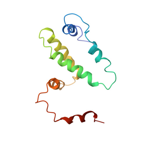
    The three-dimensional crystal structure of recombinant bovine interferon-gamma was determined using the multiple isomorphous replacement method at 3.0 A and refined to an R factor of 19.2%. This protein crystallizes in space group P2(1)2(1)2(1) with unit-cell parameters of a = 42.8, b = 79.9 and c = 85.4 A. There is one functional dimer in the asymmetric unit. The two polypeptide chains are related by a non-crystallographic twofold symmetry axis. The secondary structure is predominantly alpha-helical with extensive interdigitation of the alpha-helical segments of the polypeptide chains that make up the dimer. The secondary structure, tertiary structure and topology of this molecule are identical to the previously reported structures of recombinant rabbit interferon-gamma and recombinant human interferon-gamma. The molecular topology is also similar to that of murine interferon-beta. These structural similarities strongly indicate the presence of a unique topological feature (fold) among gamma-interferons from different species, and also among the different classes of interferons.


  • Organizational Affiliation

    National Cancer Institute-Frederick Cancer Research Development Center, ABL-Basic Research Program, Frederick, Maryland 21702, USA.


Macromolecules
Find similar proteins by:  (by identity cutoff)  |  3D Structure
Entity ID: 1
MoleculeChains Sequence LengthOrganismDetailsImage
INTERFERON-GAMMA
A, B
119Bos taurusMutation(s): 0 
UniProt
Find proteins for P07353 (Bos taurus)
Explore P07353 
Go to UniProtKB:  P07353
Entity Groups  
Sequence Clusters30% Identity50% Identity70% Identity90% Identity95% Identity100% Identity
UniProt GroupP07353
Sequence Annotations
Expand
  • Reference Sequence
Experimental Data & Validation

Experimental Data

  • Method: X-RAY DIFFRACTION
  • Resolution: 3.00 Å
  • R-Value Work: 0.192 
  • R-Value Observed: 0.192 
  • Space Group: P 21 21 21
Unit Cell:
Length ( Å )Angle ( ˚ )
a = 42.8α = 90
b = 79.9β = 90
c = 85.4γ = 90
Software Package:
Software NamePurpose
X-PLORmodel building
X-PLORrefinement
X-PLORphasing

Structure Validation

View Full Validation Report


Entry History 

Deposition Data

Revision History  (Full details and data files)

  • Version 1.0: 1994-01-31
    Type: Initial release
  • Version 1.1: 2008-03-24
    Changes: Version format compliance
  • Version 1.2: 2011-07-13
    Changes: Version format compliance
  • Version 1.3: 2024-02-14
    Changes: Data collection, Database references, Other