Prolylpeptide binding by the prokaryotic SH3-like domain of the diphtheria toxin repressor: a regulatory switch.
Wylie, G.P., Rangachari, V., Bienkiewicz, E.A., Marin, V., Bhattacharya, N., Love, J.F., Murphy, J.R., Logan, T.M.(2005) Biochemistry 44: 40-51
- PubMed: 15628844
- DOI: https://doi.org/10.1021/bi048035p
- Primary Citation of Related Structures:
1QVP, 1QW1 - PubMed Abstract:
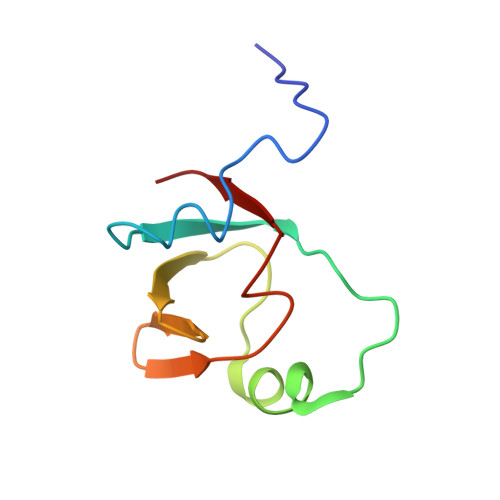
Diphtheria toxin repressor (DtxR) regulates the expression of iron-sensitive genes in Corynebacterium diphtheriae, including the diphtheria toxin gene. DtxR contains an N-terminal metal- and DNA-binding domain that is connected by a proline-rich flexible peptide segment (Pr) to a C-terminal src homology 3 (SH3)-like domain. We determined the solution structure of the intramolecular complex formed between the proline-rich segment and the SH3-like domain by use of NMR spectroscopy. The structure of the intramolecularly bound Pr segment differs from that seen in eukaryotic prolylpeptide-SH3 domain complexes. The prolylpeptide ligand is bound by the SH3-like domain in a deep crevice lined by aliphatic amino acid residues and passes through the binding site twice but does not adopt a polyprolyl type-II helix. NMR studies indicate that this intramolecular complex is present in the apo-state of the repressor. Isothermal equilibrium denaturation studies show that intramolecular complex formation contributes to the stability of the apo-repressor. The binding affinity of synthetic peptides to the SH3-like domain was determined using isothermal titration calorimetry. From the structure and the binding energies, we calculated the enhancement in binding energy for the intramolecular reaction and compared it to the energetics of dimerization. Together, the structural and biophysical studies suggest that the proline-rich peptide segment of DtxR functions as a switch that modulates the activation of repressor activity.
Organizational Affiliation:
Department of Chemistry and Biochemistry, Florida State University, Tallahassee, Florida 32306-4390, USA.


