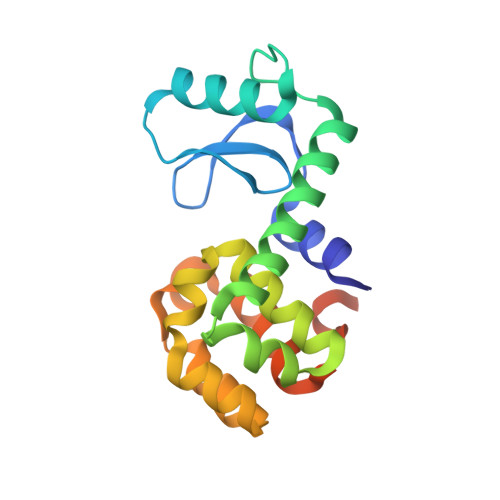
Relocation or duplication of the helix A sequence of T4 lysozyme causes only modest changes in structure but can increase or decrease the rate of folding.
Sagermann, M., Baase, W.A., Mooers, B.H., Gay, L., Matthews, B.W.(2004) Biochemistry 43: 1296-1301
- PubMed: 14756565
- DOI: https://doi.org/10.1021/bi035702q
- Primary Citation of Related Structures:
1P56, 1P5C - PubMed Abstract:
In T4 lysozyme, helix A is located at the amino terminus of the sequence but is associated with the C-terminal domain in the folded structure. To investigate the implications of this arrangement for the folding of the protein, we first created a circularly permuted variant with a new amino terminus at residue 12. In effect, this moves the sequence corresponding to helix A from the N- to the C-terminus of the molecule. The protein crystallized nonisomorphously with the wild type but has a very similar structure, showing that the unit consisting of helix A and the C-terminal domain can be reconstituted from a contiguous polypeptide chain. The protein is less stable than the wild type but folds slightly faster. We then produced a second variant in which the helix A sequence was appended at the C-terminus (as in the first variant), but was also restored at the N-terminus (as in the wild type). This variant has two helix A sequences, one at the N-terminus and the other at the C-terminus, each of which can compete for the same site in the folded protein. The crystal structure shows that it is the N-terminal sequence that folds in a manner similar to that of the wild type, whereas the copy at the C-terminus is forced to loop out. The stability of this protein is much closer to that of the wild type, but its rate of folding is significantly slower. The reduction in rate is attributed to the presence of the two identical sequence segments which compete for a single, mutually exclusive, site.
Organizational Affiliation:
Institute of Molecular Biology, Howard Hughes Medical Institute, and Department of Physics, 1229, University of Oregon, Eugene, Oregon 97403-1229, USA.


