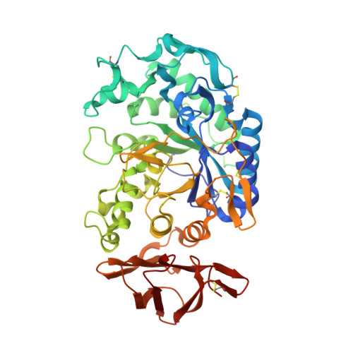
1OSE

Porcine pancreatic alpha-amylase complexed with acarbose


Experimental Data Snapshot

  • Method: X-RAY DIFFRACTION
  • Resolution: 2.30 Å
  • R-Value Free: 0.222 
  • R-Value Work: 0.176 
  • R-Value Observed: 0.176 

wwPDB Validation   3D Report Full Report


This is version 3.1 of the entry. See complete history


Literature

Crystal structure of pig pancreatic alpha-amylase isoenzyme II, in complex with the carbohydrate inhibitor acarbose.

Gilles, C.Astier, J.P.Marchis-Mouren, G.Cambillau, C.Payan, F.

(1996) Eur J Biochem 238: 561-569

  • DOI: https://doi.org/10.1111/j.1432-1033.1996.0561z.x
  • Primary Citation of Related Structures:  
    1OSE

  • PubMed Abstract: 

    Two different crystal forms of pig pancreatic alpha-amylase isoenzyme II (PPAII), free and complexed to a carbohydrate inhibitor (acarbose), have been compared together and to previously reported structures of PPAI. A crystal form obtained at 4 degrees C, containing nearly 72% solvent, made it possible to obtain a new complex with acarbose, different from a previous one obtained at 20 degrees C [Qian, M., Buisson, G., Duée, E., Haser, H. & Payan, F. (1994) Biochemistry 33, 6284-6294]. In the present form, six contiguous subsites of the enzyme active site are occupied by the carbohydrate ligand; the structural data indicate that the binding site is capable of holding more than the five glucose units of the scheme proposed through kinetic studies. A monosaccharide ring bridging two protein molecules related by the crystal packing is located on the surface, at a distance of 2.0 nm from the reducing end of the inhibitor ligand; the symmetry-related glucose ring in the crystal lattice is found 1.5 nm away from the non-reducing end of the inhibitor ligand.


  • Organizational Affiliation

    LCCMB-IBSM, CNRS, Marseille, France.


Macromolecules
Find similar proteins by:  (by identity cutoff)  |  3D Structure
Entity ID: 1
MoleculeChains Sequence LengthOrganismDetailsImage
PORCINE ALPHA-AMYLASE496Sus scrofaMutation(s): 0 
EC: 3.2.1.1
UniProt
Find proteins for P00690 (Sus scrofa)
Explore P00690 
Go to UniProtKB:  P00690
Entity Groups  
Sequence Clusters30% Identity50% Identity70% Identity90% Identity95% Identity100% Identity
UniProt GroupP00690
Sequence Annotations
Expand
  • Reference Sequence
Oligosaccharides

Help

Entity ID: 2
MoleculeChains Length2D Diagram Glycosylation3D Interactions
4,6-dideoxy-4-{[(1S,4R,5S,6S)-4,5,6-trihydroxy-3-(hydroxymethyl)cyclohex-2-en-1-yl]amino}-alpha-D-glucopyranose-(1-4)-alpha-D-glucopyranose-(1-4)-4,6-dideoxy-4-{[(1S,4R,5S,6S)-4,5,6-trihydroxy-3-(hydroxymethyl)cyclohex-2-en-1-yl]amino}-alpha-D-glucopyranose-(1-4)-beta-D-glucopyranose
B
4N/AN/A
Glycosylation Resources
GlyTouCan:  G94545JA
GlyCosmos:  G94545JA
Small Molecules
Modified Residues  1 Unique
IDChains TypeFormula2D DiagramParent
PCA
Query on PCA
A
L-PEPTIDE LINKINGC5 H7 N O3GLN
Experimental Data & Validation

Experimental Data

  • Method: X-RAY DIFFRACTION
  • Resolution: 2.30 Å
  • R-Value Free: 0.222 
  • R-Value Work: 0.176 
  • R-Value Observed: 0.176 
  • Space Group: P 21 21 21
Unit Cell:
Length ( Å )Angle ( ˚ )
a = 70.6α = 90
b = 114.7β = 90
c = 118.5γ = 90
Software Package:
Software NamePurpose
XDSdata scaling
X-PLORmodel building
X-PLORrefinement
XDSdata reduction
X-PLORphasing

Structure Validation

View Full Validation Report


Entry History 

Deposition Data

Revision History  (Full details and data files)

  • Version 1.0: 1997-04-01
    Type: Initial release
  • Version 1.1: 2008-03-24
    Changes: Version format compliance
  • Version 1.2: 2011-07-13
    Changes: Version format compliance
  • Version 2.0: 2019-12-25
    Changes: Data collection, Derived calculations, Polymer sequence
  • Version 3.0: 2020-07-29
    Type: Remediation
    Reason: Carbohydrate remediation
    Changes: Atomic model, Data collection, Derived calculations, Structure summary
  • Version 3.1: 2024-10-09
    Changes: Data collection, Database references, Derived calculations, Structure summary