1OAG

Ascorbate peroxidase from soybean cytosol


Experimental Data Snapshot

  • Method: X-RAY DIFFRACTION
  • Resolution: 1.75 Å
  • R-Value Free: 0.177 
  • R-Value Work: 0.142 
  • R-Value Observed: 0.144 

Starting Model: experimental
View more details

wwPDB Validation   3D Report Full Report


Ligand Structure Quality Assessment 


This is version 1.4 of the entry. See complete history


Literature

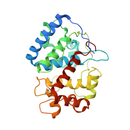
Crystal Structure of the Ascorbate Peroxidase-Ascorbate Complex

Sharp, K.H.Mewies, M.Moody, P.C.E.Raven, E.L.

(2003) Nat Struct Biol 10: 303

  • DOI: https://doi.org/10.1038/nsb913
  • Primary Citation of Related Structures:  
    1OAF, 1OAG

  • PubMed Abstract: 

    Heme peroxidases catalyze the H2O2-dependent oxidation of a variety of substrates, most of which are organic. Mechanistically, these enzymes are well characterized: they share a common catalytic cycle that involves formation of a two-electron, oxidized Compound I intermediate followed by two single-electron reduction steps by substrate. The substrate specificity is more diverse--most peroxidases oxidize small organic substrates, but there are prominent exceptions--and there is a notable absence of structural information for a representative peroxidase-substrate complex. Thus, the features that control substrate specificity remain undefined. We present the structure of the complex of ascorbate peroxidase-ascorbate. The structure defines the ascorbate-binding interaction for the first time and provides new rationalization of the unusual functional features of the related cytochrome c peroxidase enzyme, which has been a benchmark for peroxidase catalysis for more than 20 years. A new mechanism for electron transfer is proposed that challenges existing views of substrate oxidation in other peroxidases.


  • Organizational Affiliation

    Department of Chemistry, University of Leicester, University Road, Leicester, LE1 7RH, England, UK.


Macromolecules
Find similar proteins by:  (by identity cutoff)  |  3D Structure
Entity ID: 1
MoleculeChains Sequence LengthOrganismDetailsImage
ASCORBATE PEROXIDASE261Glycine maxMutation(s): 0 
EC: 1.11.1.11
UniProt
Find proteins for Q43758 (Glycine max)
Explore Q43758 
Go to UniProtKB:  Q43758
Entity Groups  
Sequence Clusters30% Identity50% Identity70% Identity90% Identity95% Identity100% Identity
UniProt GroupQ43758
Sequence Annotations
Expand
  • Reference Sequence
Small Molecules
Ligands 2 Unique
IDChains Name / Formula / InChI Key2D Diagram3D Interactions
HEM
Query on HEM

Download Ideal Coordinates CCD File 
B [auth A]PROTOPORPHYRIN IX CONTAINING FE
C34 H32 Fe N4 O4
KABFMIBPWCXCRK-RGGAHWMASA-L
SO4
Query on SO4

Download Ideal Coordinates CCD File 
C [auth A]SULFATE ION
O4 S
QAOWNCQODCNURD-UHFFFAOYSA-L
Experimental Data & Validation

Experimental Data

  • Method: X-RAY DIFFRACTION
  • Resolution: 1.75 Å
  • R-Value Free: 0.177 
  • R-Value Work: 0.142 
  • R-Value Observed: 0.144 
  • Space Group: P 42 21 2
Unit Cell:
Length ( Å )Angle ( ˚ )
a = 81.842α = 90
b = 81.842β = 90
c = 75.031γ = 90
Software Package:
Software NamePurpose
REFMACrefinement
DENZOdata reduction
SCALEPACKdata scaling
MOLREPphasing

Structure Validation

View Full Validation Report


Ligand Structure Quality Assessment 


Entry History 

Deposition Data

Revision History  (Full details and data files)

  • Version 1.0: 2003-03-20
    Type: Initial release
  • Version 1.1: 2011-05-08
    Changes: Version format compliance
  • Version 1.2: 2011-07-13
    Changes: Version format compliance
  • Version 1.3: 2019-07-24
    Changes: Data collection, Derived calculations
  • Version 1.4: 2023-12-13
    Changes: Data collection, Database references, Derived calculations, Other, Refinement description