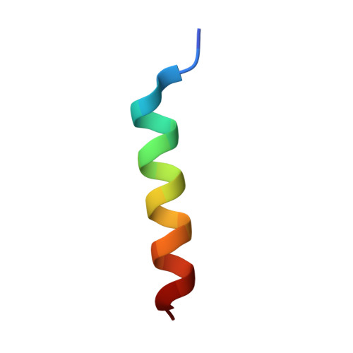
Structure and ubiquitin binding of the ubiquitin-interacting motif.
Fisher, R.D., Wang, B., Alam, S.L., Higginson, D.S., Robinson, H., Sundquist, W.I., Hill, C.P.(2003) J Biol Chem 278: 28976-28984
- PubMed: 12750381
- DOI: https://doi.org/10.1074/jbc.M302596200
- Primary Citation of Related Structures:
1O06 - PubMed Abstract:
Ubiquitylation is used to target proteins into a large number of different biological processes including proteasomal degradation, endocytosis, virus budding, and vacuolar protein sorting (Vps). Ubiquitylated proteins are typically recognized using one of several different conserved ubiquitin binding modules. Here, we report the crystal structure and ubiquitin binding properties of one such module, the ubiquitin-interacting motif (UIM). We found that UIM peptides from several proteins involved in endocytosis and vacuolar protein sorting including Hrs, Vps27p, Stam1, and Eps15 bound specifically, but with modest affinity (Kd = 0.1-1 mm), to free ubiquitin. Full affinity ubiquitin binding required the presence of conserved acidic patches at the N and C terminus of the UIM, as well as highly conserved central alanine and serine residues. NMR chemical shift perturbation mapping experiments demonstrated that all of these UIM peptides bind to the I44 surface of ubiquitin. The 1.45 A resolution crystal structure of the second yeast Vps27p UIM (Vps27p-2) revealed that the ubiquitin-interacting motif forms an amphipathic helix. Although Vps27p-2 is monomeric in solution, the motif unexpectedly crystallized as an antiparallel four-helix bundle, and the potential biological implications of UIM oligomerization are therefore discussed.
Organizational Affiliation:
Department of Biochemistry, University of Utah School of Medicine, Salt Lake City, Utah 84132, USA.


