1LWS

Crystal structure of the intein homing endonuclease PI-SceI bound to its recognition sequence


Experimental Data Snapshot

  • Method: X-RAY DIFFRACTION
  • Resolution: 3.50 Å
  • R-Value Free: 0.310 
  • R-Value Work: 0.287 

wwPDB Validation   3D Report Full Report


This is version 1.2 of the entry. See complete history


Literature

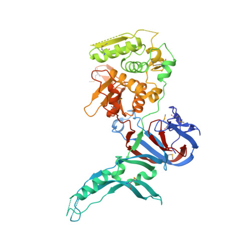
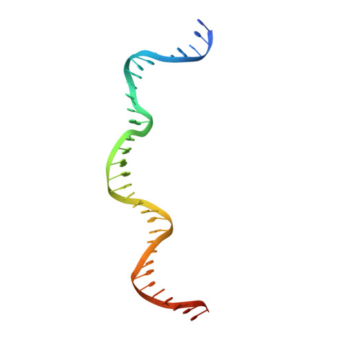
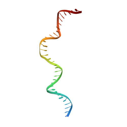
Crystal structure of the intein homing endonuclease PI-SceI bound to its recognition sequence.

Moure, C.M.Gimble, F.S.Quiocho, F.A.

(2002) Nat Struct Biol 9: 764-770

  • DOI: https://doi.org/10.1038/nsb840
  • Primary Citation of Related Structures:  
    1LWS, 1LWT

  • PubMed Abstract: 

    The first X-ray structures of an intein-DNA complex, that of the two-domain homing endonuclease PI-SceI bound to its 36-base pair DNA substrate, have been determined in the presence and absence of Ca(2+). The DNA shows an asymmetric bending pattern, with a major 50 degree bend in the endonuclease domain and a minor 22 degree bend in the splicing domain region. Distortions of the DNA bound to the endonuclease domain cause the insertion of the two cleavage sites in the catalytic center. DNA binding induces changes in the protein conformation. The two overlapping non-identical active sites in the endonucleolytic center contain two Ca(+2) ions that coordinate to the catalytic Asp residues. Structure analysis indicates that the top strand may be cleaved first.


  • Organizational Affiliation

    Howard Hughes Medical Institute, Baylor College of Medicine, Houston, Texas 77030, USA.


Macromolecules

Find similar proteins by:  (by identity cutoff)  |  3D Structure
Entity ID: 3
MoleculeChains Sequence LengthOrganismDetailsImage
ENDONUCLEASE PI-SCEIC [auth A]454Saccharomyces cerevisiaeMutation(s): 8 
Gene Names: VMA1
EC: 3.1
UniProt
Find proteins for P17255 (Saccharomyces cerevisiae (strain ATCC 204508 / S288c))
Explore P17255 
Go to UniProtKB:  P17255
Entity Groups  
Sequence Clusters30% Identity50% Identity70% Identity90% Identity95% Identity100% Identity
UniProt GroupP17255
Sequence Annotations
Expand
  • Reference Sequence
Find similar nucleic acids by:  (by identity cutoff)  |  3D Structure
Entity ID: 1
MoleculeChains LengthOrganismImage
PI-SceI DNA recognition region top strandA [auth B]37N/A
Sequence Annotations
Expand
  • Reference Sequence
Find similar nucleic acids by:  (by identity cutoff)  |  3D Structure
Entity ID: 2
MoleculeChains LengthOrganismImage
PI-SceI DNA recognition region bottom strandB [auth C]37N/A
Sequence Annotations
Expand
  • Reference Sequence
Small Molecules
Modified Residues  1 Unique
IDChains TypeFormula2D DiagramParent
MSE
Query on MSE
C [auth A]L-PEPTIDE LINKINGC5 H11 N O2 SeMET
Experimental Data & Validation

Experimental Data

  • Method: X-RAY DIFFRACTION
  • Resolution: 3.50 Å
  • R-Value Free: 0.310 
  • R-Value Work: 0.287 
  • Space Group: P 65 2 2
Unit Cell:
Length ( Å )Angle ( ˚ )
a = 123α = 90
b = 123β = 90
c = 211.7γ = 120
Software Package:
Software NamePurpose
DENZOdata reduction
SCALEPACKdata scaling
CNSrefinement
CNSphasing

Structure Validation

View Full Validation Report


Entry History 

Deposition Data

Revision History  (Full details and data files)

  • Version 1.0: 2002-09-27
    Type: Initial release
  • Version 1.1: 2008-04-28
    Changes: Version format compliance
  • Version 1.2: 2011-07-13
    Changes: Version format compliance